| HepG2 | 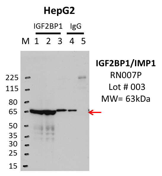
Caption: IP-WB analysis of HepG2 whole cell lysate using IGF2BP1 specific antibody. Lane 1 is 1% twenty million whole cell lysate input, lane 2 is 1% of the supernatant afer IP and Lane 3 is 10% of IP enrichment using rabbit polyclonal anti-IGF2BP1 Antibody. Lane 4 is 1% ten million whole cell lysate input and lane 5 is 25% IP enrichment using rabbit normal IgG (lanes under 'IgG'). Method: immunoprecipitation Releated Sample: eCLIP:205 Status: Released Lab: Yeo Lab | 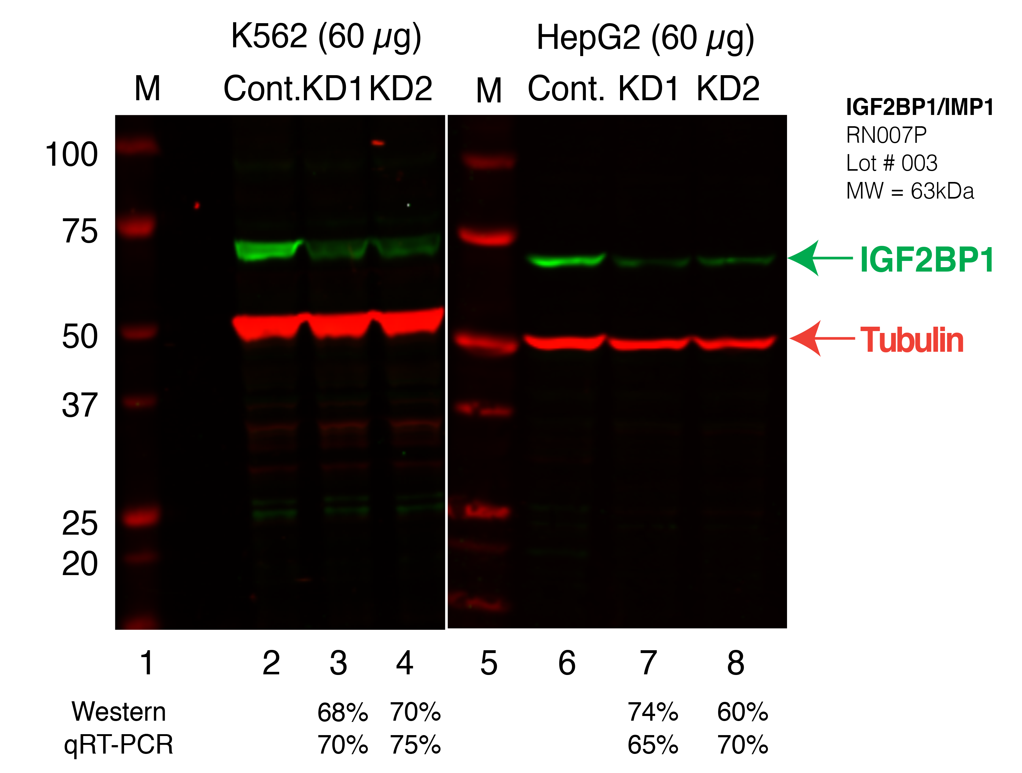
Caption: Western blot following shRNA against IGF2BP1 in K562 and HepG2 whole cell lysate using IGF2BP1 specific antibody. Lane 1 is a ladder, lane 2 is K562 non-targeting control knockdown, lane 3 and 4 are two different shRNAs against IGF2BP1. Lanes 5-8 follow the same pattern, but in HepG2. IGF2BP1 protein appears as the green band, Tubulin serves as a control and appears in red. Releated Sample: BGKLV11-17 Status: Released Lab: Graveley Lab 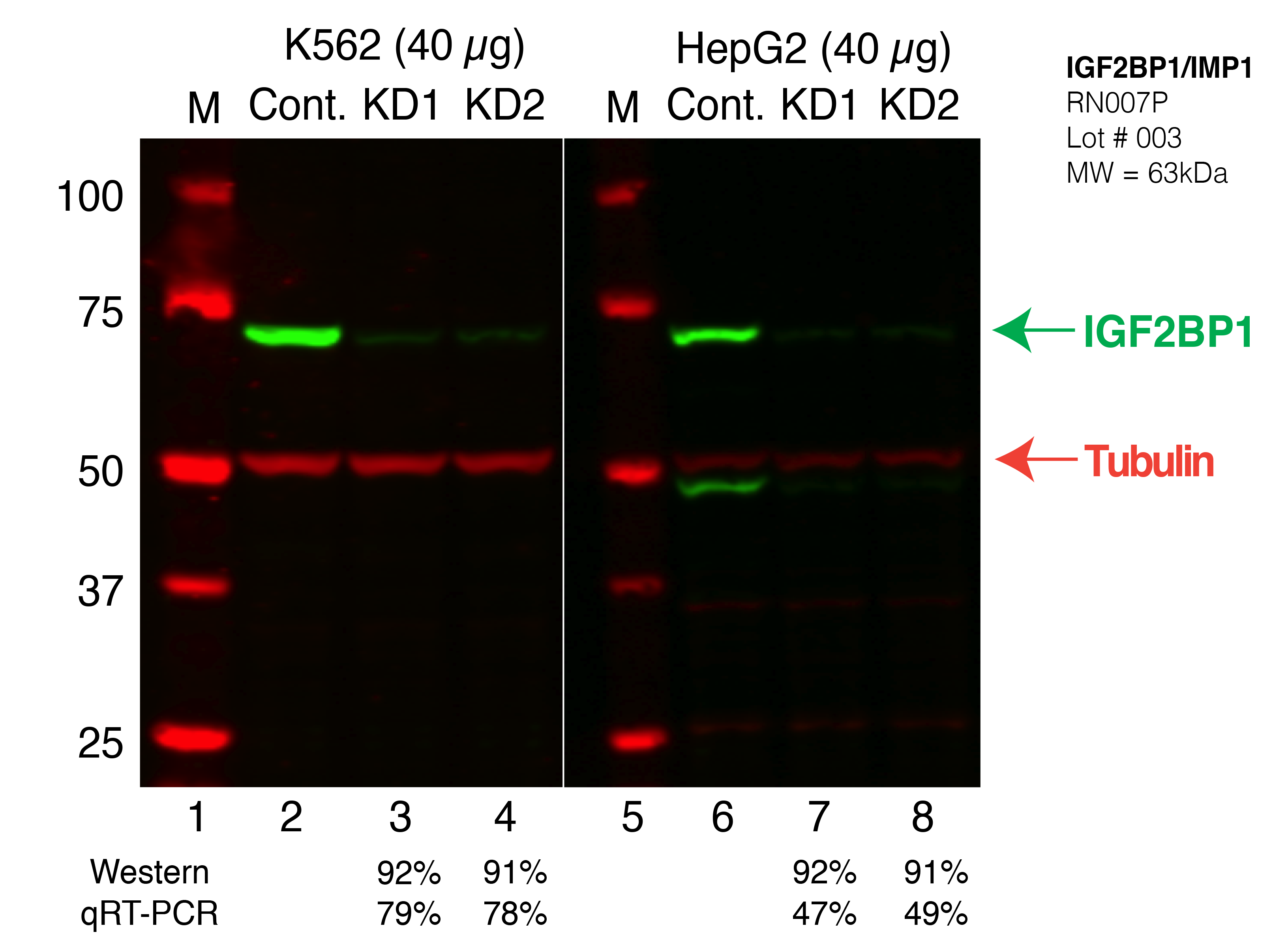
Caption: Western blot following CRISPR against IGF2BP1 in K562 and HepG2 whole cell lysate using IGF2BP1 specific antibody. Lane 1 is a ladder, lane 2 is K562 non-targeting control knockdown, lane 3 and 4 are two different CRISPR against IGF2BP1. 5-8 follow the same pattern, but in HepG2. IGF2BP1 protein appears as the green band, Tubulin serves as a control and appears in red. Releated Sample: BGKcLV02-3 Status: Released Lab: Graveley Lab |
|---|
| K562 | 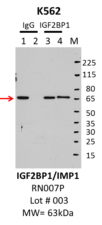
Caption: IP-Western Blot analysis of K562 whole cell lysate using IGF2BP1 specific antibody. Lane 1 is 2.5% of five million whole cell lysate input, lane 2 is 2.5% of supernatant after immunoprecipitation and Lane 3 is 50% of IP enrichment using either rabbit polyclonal anti-IGF2BP1 antibody (lanes under 'IGF2BP1') or using rabbit normal IgG (lanes under 'IgG'). Method: immunoprecipitation Releated Sample: eCLIP:220 Status: Released Lab: Yeo Lab |
Caption: Western blot following shRNA against IGF2BP1 in K562 and HepG2 whole cell lysate using IGF2BP1 specific antibody. Lane 1 is a ladder, lane 2 is K562 non-targeting control knockdown, lane 3 and 4 are two different shRNAs against IGF2BP1. Lanes 5-8 follow the same pattern, but in HepG2. IGF2BP1 protein appears as the green band, Tubulin serves as a control and appears in red. Releated Sample: BGKLV11-17 Status: Released Lab: Graveley Lab
Caption: Western blot following CRISPR against IGF2BP1 in K562 and HepG2 whole cell lysate using IGF2BP1 specific antibody. Lane 1 is a ladder, lane 2 is K562 non-targeting control knockdown, lane 3 and 4 are two different CRISPR against IGF2BP1. 5-8 follow the same pattern, but in HepG2. IGF2BP1 protein appears as the green band, Tubulin serves as a control and appears in red. Releated Sample: BGKcLV02-3 Status: Released Lab: Graveley Lab |
|---|