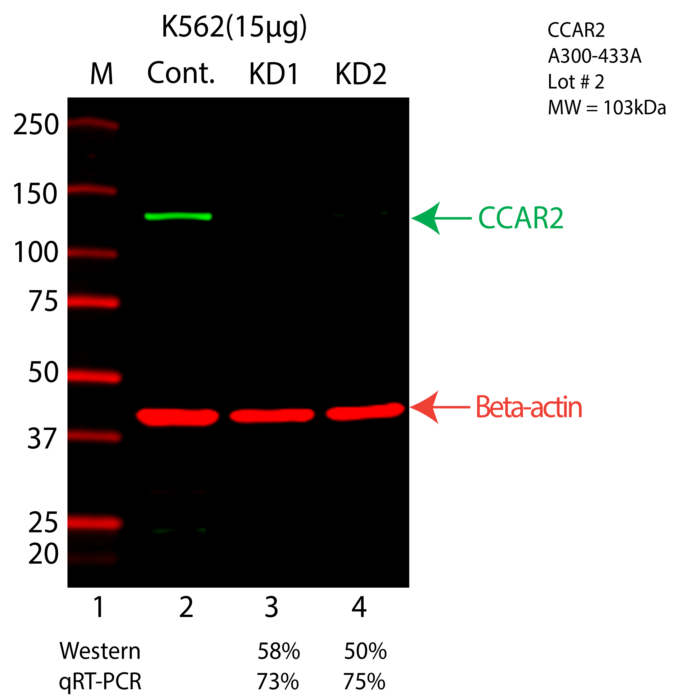
| CCAR2 | Product_ID: A300-433A
Lot_ID: 2
Source: Bethyl Labs
Target Name: CCAR2-human | | | 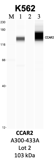
| 
| | |
|---|
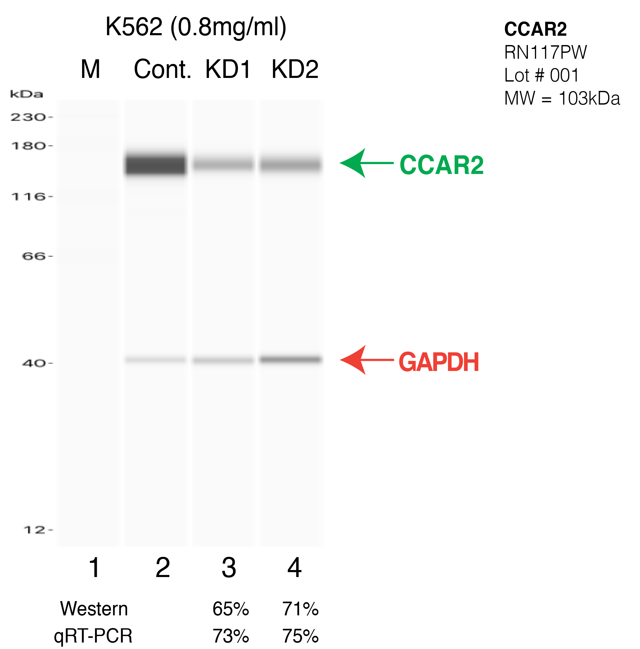
| CCAR2 | Product_ID: RN117PW
Lot_ID: 1
Source: MBLI
Target Name: CCAR2-human | | | 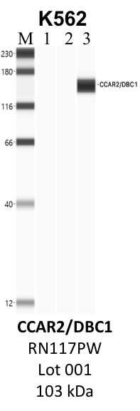
| 
| | |
|---|
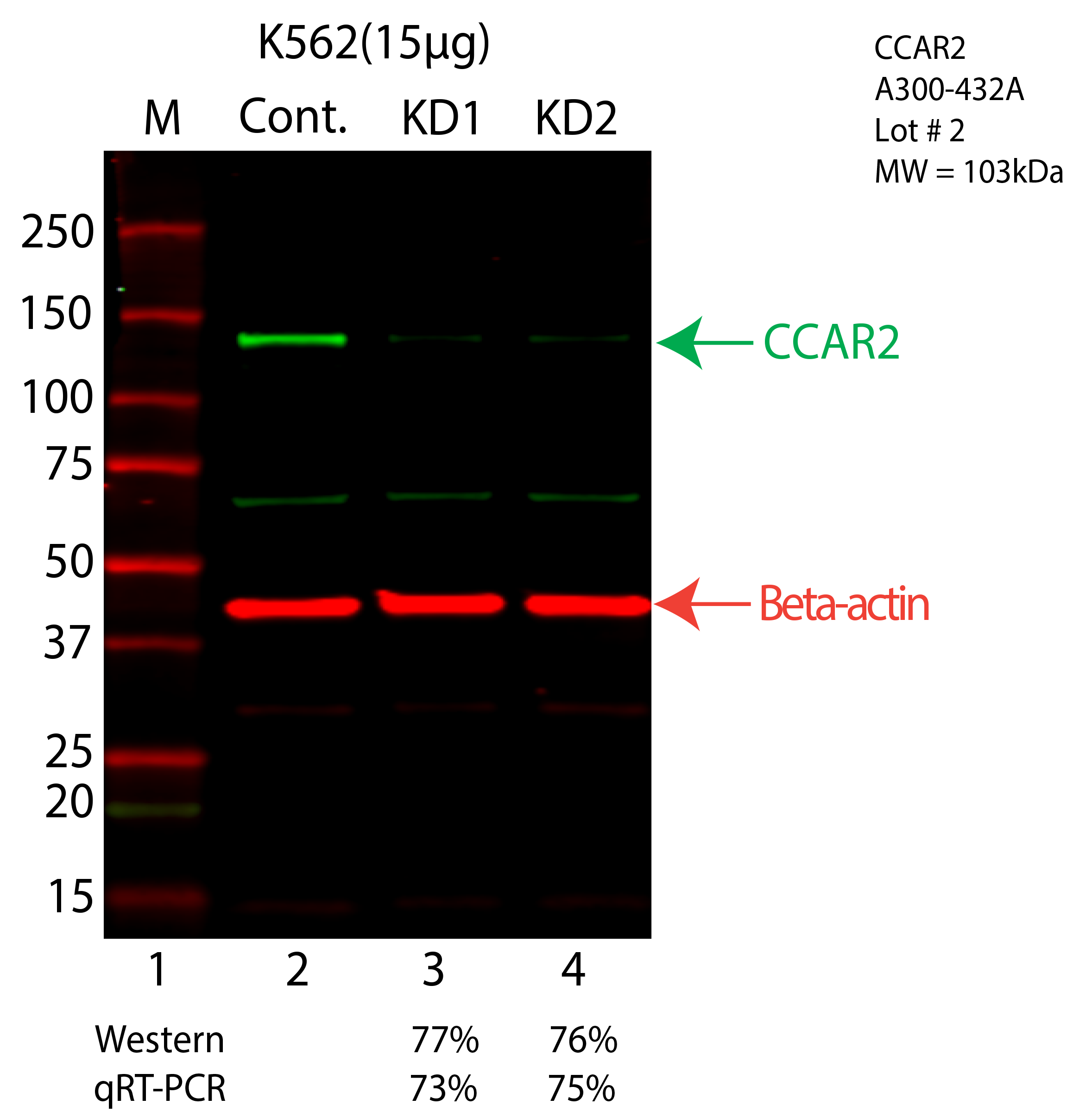
| CCAR2 | Product_ID: A300-432A
Lot_ID: 2
Source: Bethyl Labs
Target Name: CCAR2-human | | | 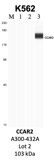
| 
| | |
|---|
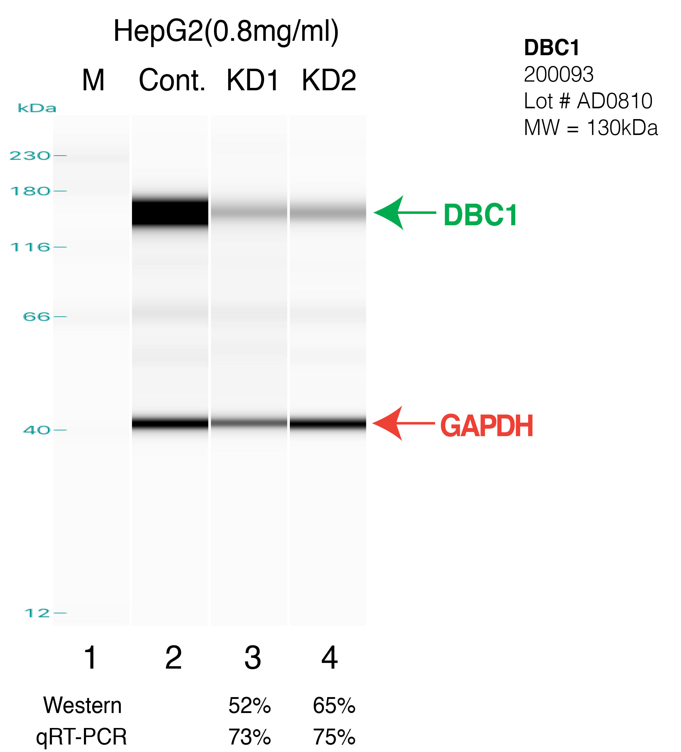
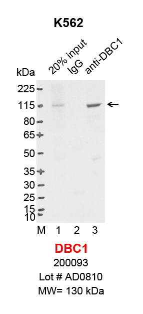
| CCAR2 | Product_ID: 200093
Lot_ID: AD0810
Source: Zen Bioscience
Target Name: CCAR2-human | 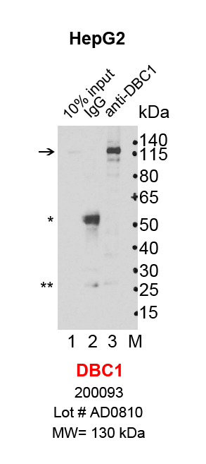
| 
| 
| | | |
|---|
| CCAR2 | Product_ID: A303-942A
Lot_ID: 1
Source: Bethyl Labs
Target Name: CCAR2-human | | 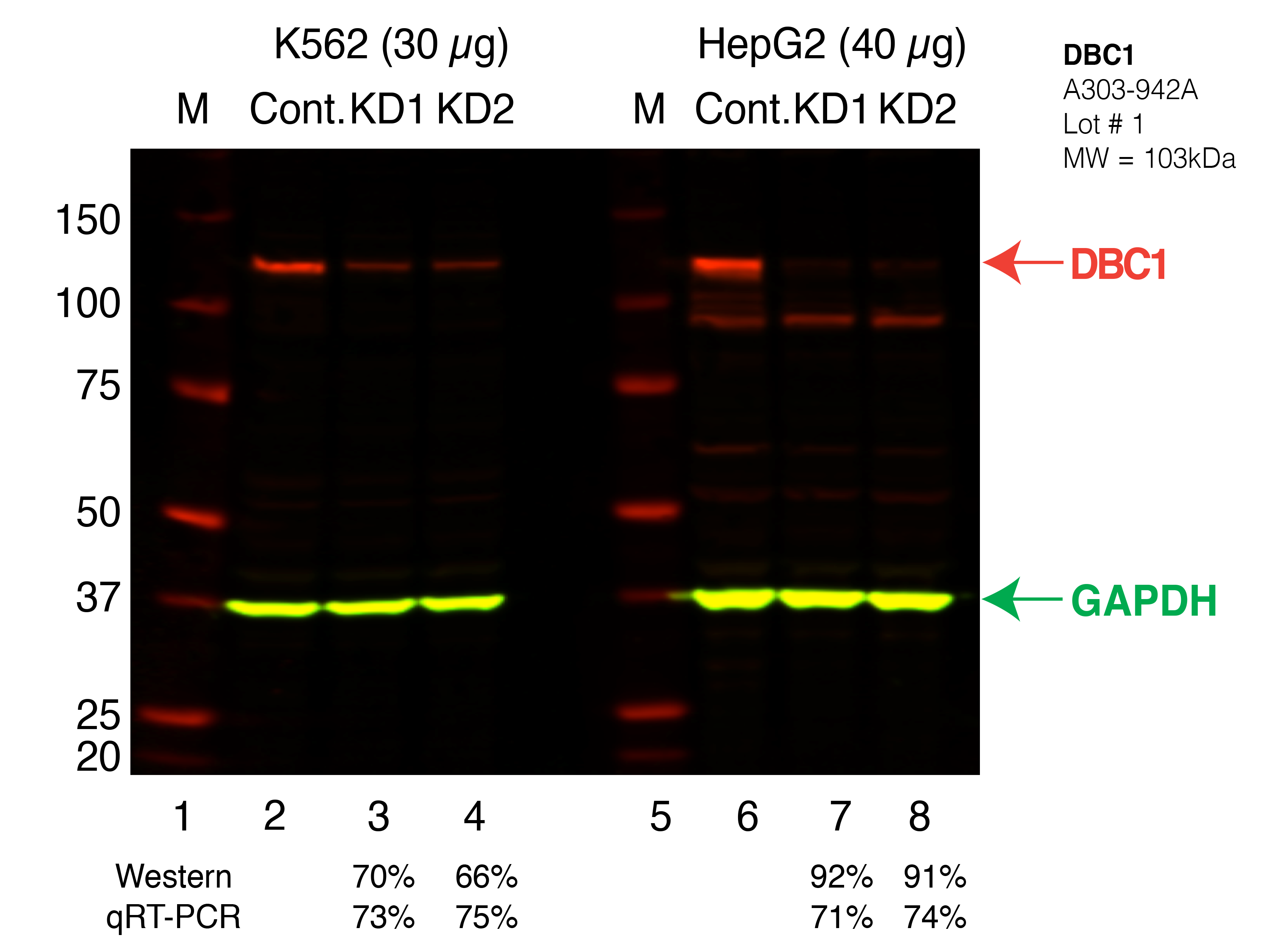
| |
| | |
|---|