| HepG2 | 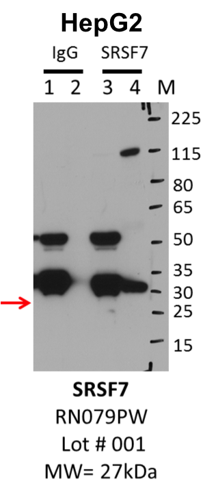
Caption: IP-Western Blot analysis of HepG2 whole cell lysate using SRSF7 specific antibody. Lane 1 is 1% of twenty million whole cell lysate input and lane 2 is 25% of IP enrichment using rabbit normal IgG (lanes under 'IgG'). Lane 3 is 1% of twenty million whole cell lysate input and lane 4 is 10% IP enrichment using rabbit polyclonal anti-SRSF7 antibody (lanes under 'SRSF7'). Method: immunoprecipitation Releated Sample: eCLIP:209 Status: Released Lab: Yeo Lab | 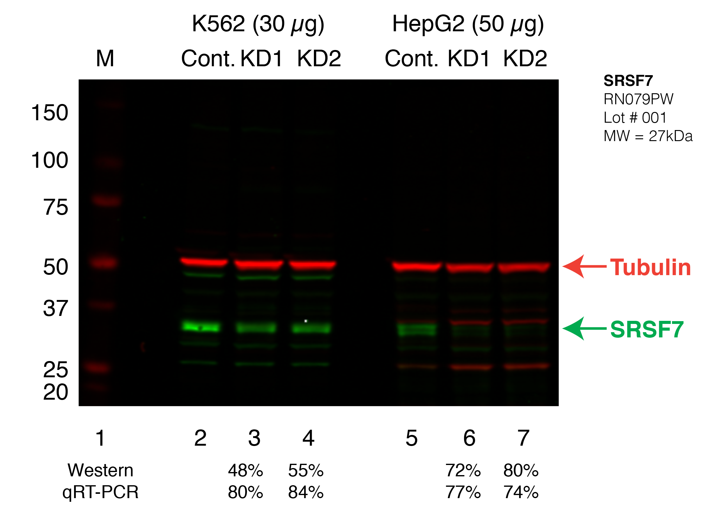
Caption: Western blot following shRNA against SRSF7 in K562 and HepG2 whole cell lysate using SRSF7 specific antibody. Lane 1 is a ladder, lane 2 is K562 non-targeting control knockdown, lane 2 and 3 are two different shRNAs against SRSF7. Lanes 5-7 follow the same pattern, but in HepG2. SRSF7 protein appears as the green band, GAPDH serves as a control and appears in red. Releated Sample: BGKLV11-53 Status: Released Lab: Graveley Lab 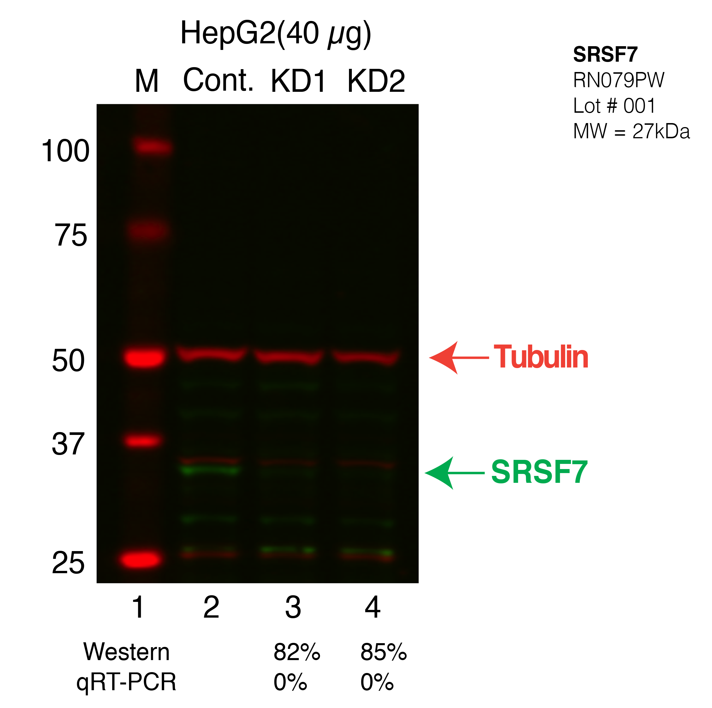
Caption: Western blot following CRISPR against SRSF7 in HepG2 whole cell lysate using SRSF7 specific antibody. Lane 1 is a ladder, lane 2 is HepG2 non-targeting control knockdown, lane 3 and 4 are two different CRISPR against SRSF7. SRSF7 protein appears as the green band, Tubulin serves as a control and appears in red. Releated Sample: BGHcLV03-5 Status: Released Lab: Graveley Lab |
|---|
| K562 | 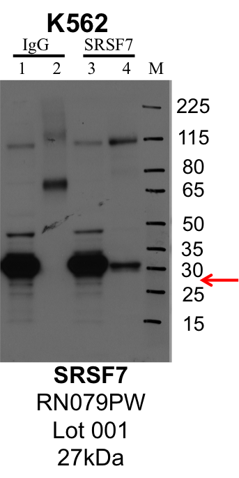
Caption: IP-Western Blot analysis of K562 whole cell lysate using SRSF7 specific antibody. Lane 1 is 1% of twenty million whole cell lysate input and lane 2 is 25% of IP enrichment using rabbit normal IgG (lanes under 'IgG'). Lane 3 is 1% of twenty million whole cell lysate input and lane 4 is 10% IP enrichment using rabbit polyclonal anti-SRSF7 antibody (lanes under 'SRSF7'). Method: immunoprecipitation Releated Sample: eCLIP:376 Status: Released Lab: Yeo Lab |
Caption: Western blot following shRNA against SRSF7 in K562 and HepG2 whole cell lysate using SRSF7 specific antibody. Lane 1 is a ladder, lane 2 is K562 non-targeting control knockdown, lane 2 and 3 are two different shRNAs against SRSF7. Lanes 5-7 follow the same pattern, but in HepG2. SRSF7 protein appears as the green band, GAPDH serves as a control and appears in red. Releated Sample: BGKLV11-53 Status: Released Lab: Graveley Lab 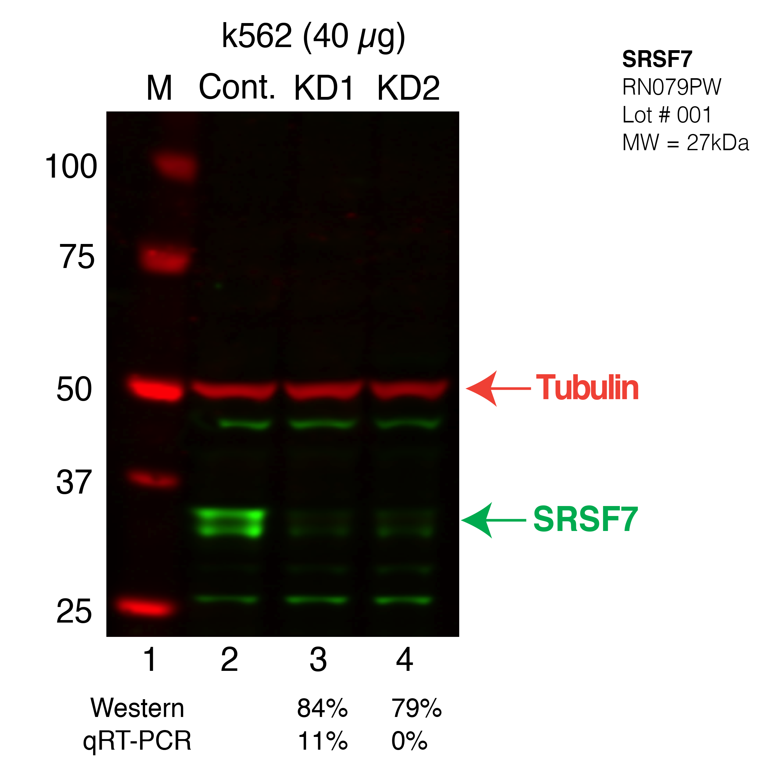
Caption: Western blot following CRISPR against SRSF7 in k562 whole cell lysate using SRSF7 specific antibody. Lane 1 is a ladder, lane 2 is k562 non-targeting control knockdown, lane 3 and 4 are two different CRISPR against SRSF7. SRSF7 protein appears as the green band, Tubulin serves as a control and appears in red. Releated Sample: BGKcLV02-5 Status: Released Lab: Graveley Lab |
|---|