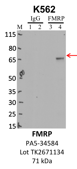
Caption: IP-WB analysis of K562 whole cell lysate using the FMR1 specific antibody, PA5-34584. Lanes 1 and 3 are 2.5% of five million whole cell lysate input. Lanes 2 and 4 are 50% of IP enrichment from five million whole cell lysate using normal IgG antibody and the FMR1-specific antibody, PA5-34584. The same antibody was used to detect protein levels via Western blot. This antibody passes preliminary validation and will be further pursued for secondary validation. *NOTE* Protein sizes are taken from Genecards.org and are only estimates based on sequence. Actual protein size may differ based on protein characteristics and electrophoresis method used. Method: immunoprecipitation Releated Sample: eCLIP:236, eCLIP:4030 Status: Released Lab: Yeo Lab |