| HepG2 | | 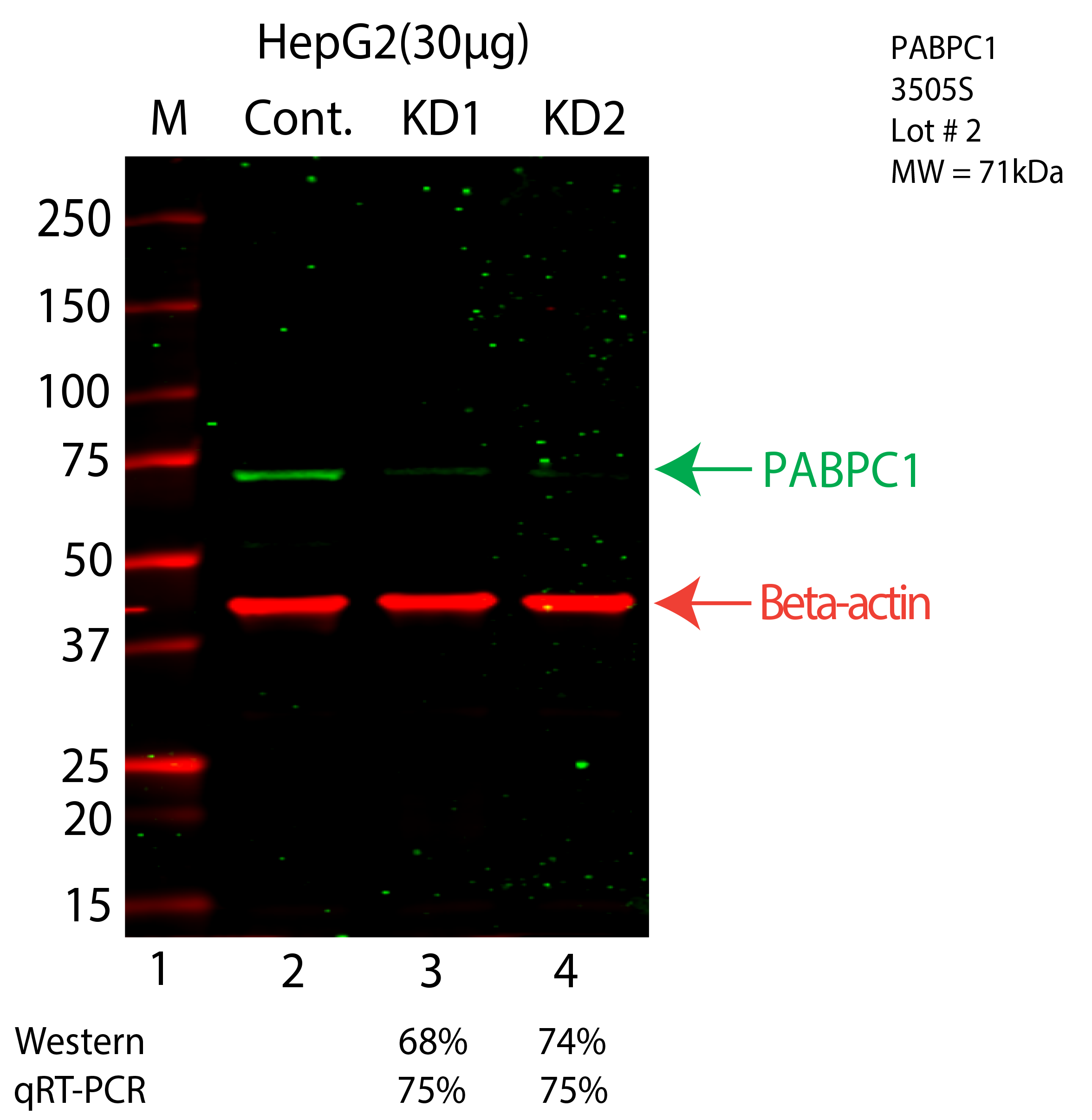
Caption: Western blot following CRISPR against PABPC1 in HepG2 whole cell lysate using PABPC1 specific antibody. Lane 1 is a ladder, lane 2 is HepG2 non-targeting control knockdown, lane 3 and 4 are two different CRISPR against PABPC1. PABPC1 protein appears as the green arrow, Beta-actin serves as a control and appears in red arrow. Releated Sample: BGHcLV43-37 Status: Submitted Lab: Graveley Lab |
|---|
| K562 | 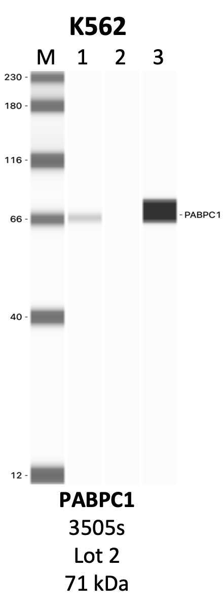
Caption: IP-WB analysis of 3505S whole cell lysate using the PABPC1 specific antibody, 3505S. Lanes 1 and 2 are 2.5% of five million whole cell lysate input and 50% of IP enrichment, respectively, using a normal IgG antibody. Lane 3 is 50% of IP enrichment from five million whole cell lysate using the PABPC1-specific antibody, 3505S. The same antibody was used to detect protein levels via Western blot. This antibody passes preliminary validation and will be further pursued for secondary validation. *NOTE* Protein sizes are taken from Genecards.org and are only estimates based on sequence. Actual protein size may differ based on protein characteristics and electrophoresis method used. Method: immunoprecipitation Status: Submitted Lab: Yeo Lab | 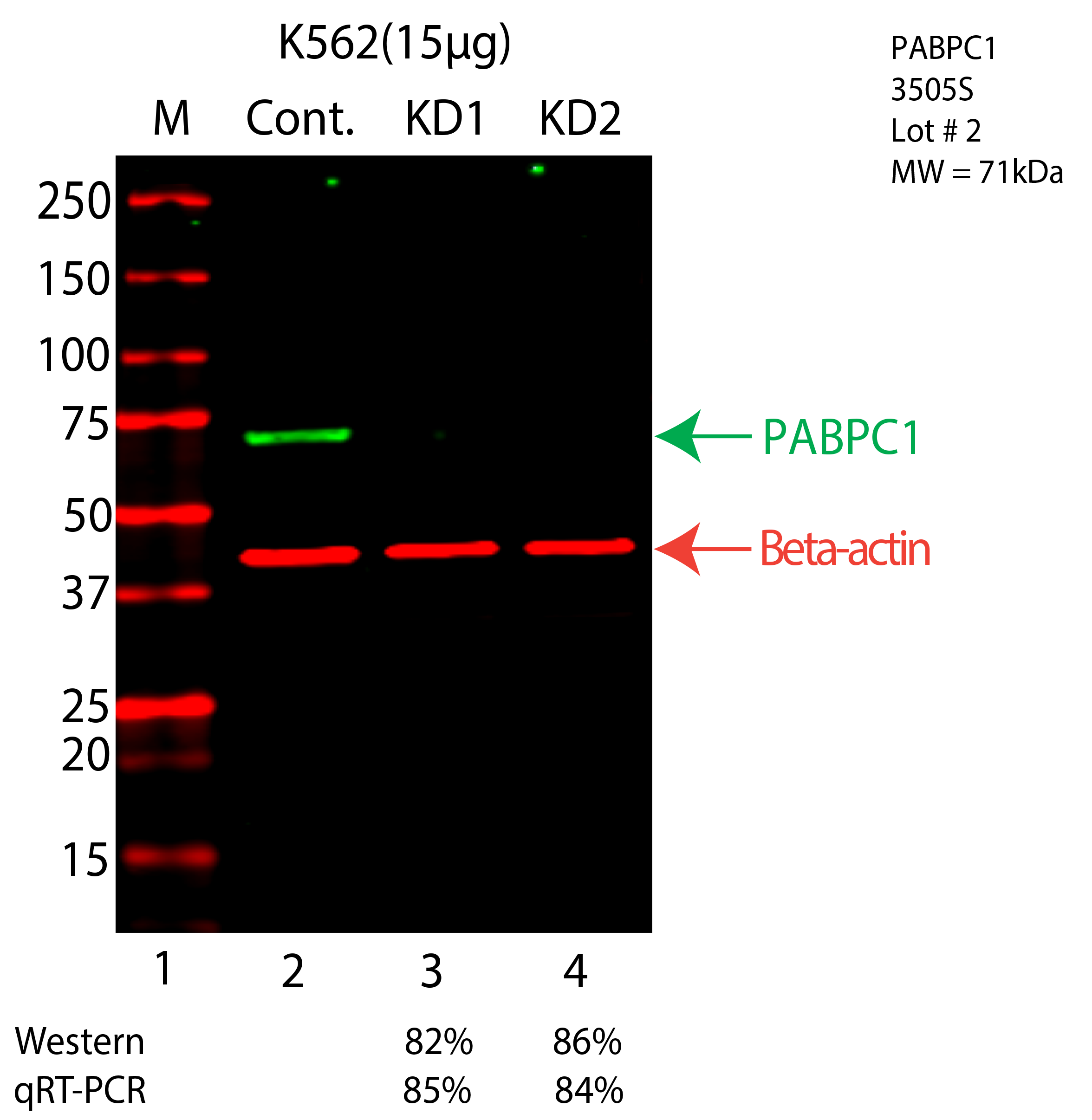
Caption: Western blot following CRISPR against PABPC1 in K562 whole cell lysate using PABPC1 specific antibody. Lane 1 is a ladder, lane 2 is K562 non-targeting control knockdown, lane 3 and 4 are two different CRISPR against PABPC1. PABPC1 protein appears as the green arrow, Beta-actin serves as a control and appears in red arrow. Releated Sample: BGKcLV42-61 Status: Submitted Lab: Graveley Lab |
|---|