Home
NEWS
Summary
Data Files
Search
FAQ
Admin Login
Top
Antibodies of SF1
3
antibodies are included.
Released
NotSatisfied
Submitted
NotReviewed
OnHold
RequestToSubmit
Experimented
RBP
Antibody Info
Primary-HepG2
Secondary-HepG2
Primary-K562
Secondary-K562
Primary-UBERON
Primary-Hela
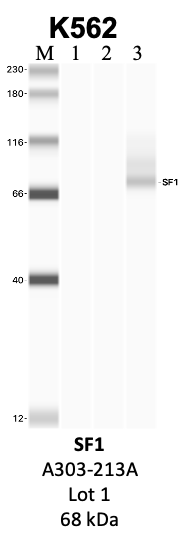
SF1
Product_ID:
A303-213A
Lot_ID: 1
Source: Bethyl Labs
Target Name: SF1-human
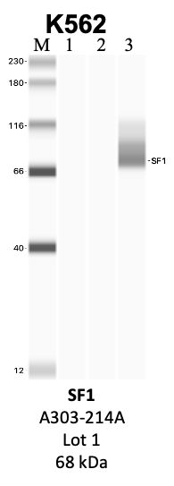
SF1
Product_ID:
A303-214A
Lot_ID: 1
Source: Bethyl Labs
Target Name: SF1-human
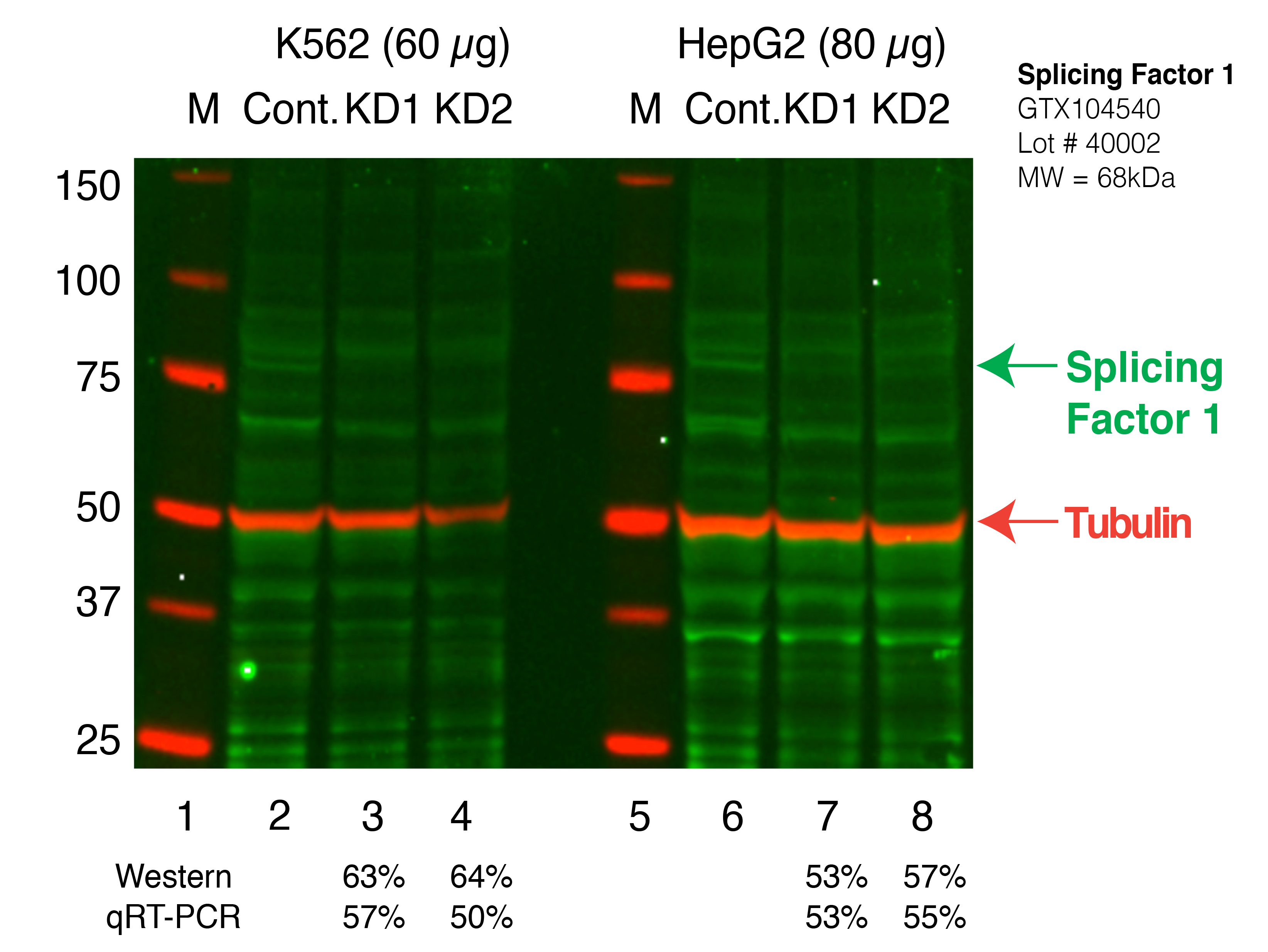
SF1
Product_ID:
GTX104540
Lot_ID: 40002
Source: GeneTex
Target Name: SF1-human
×