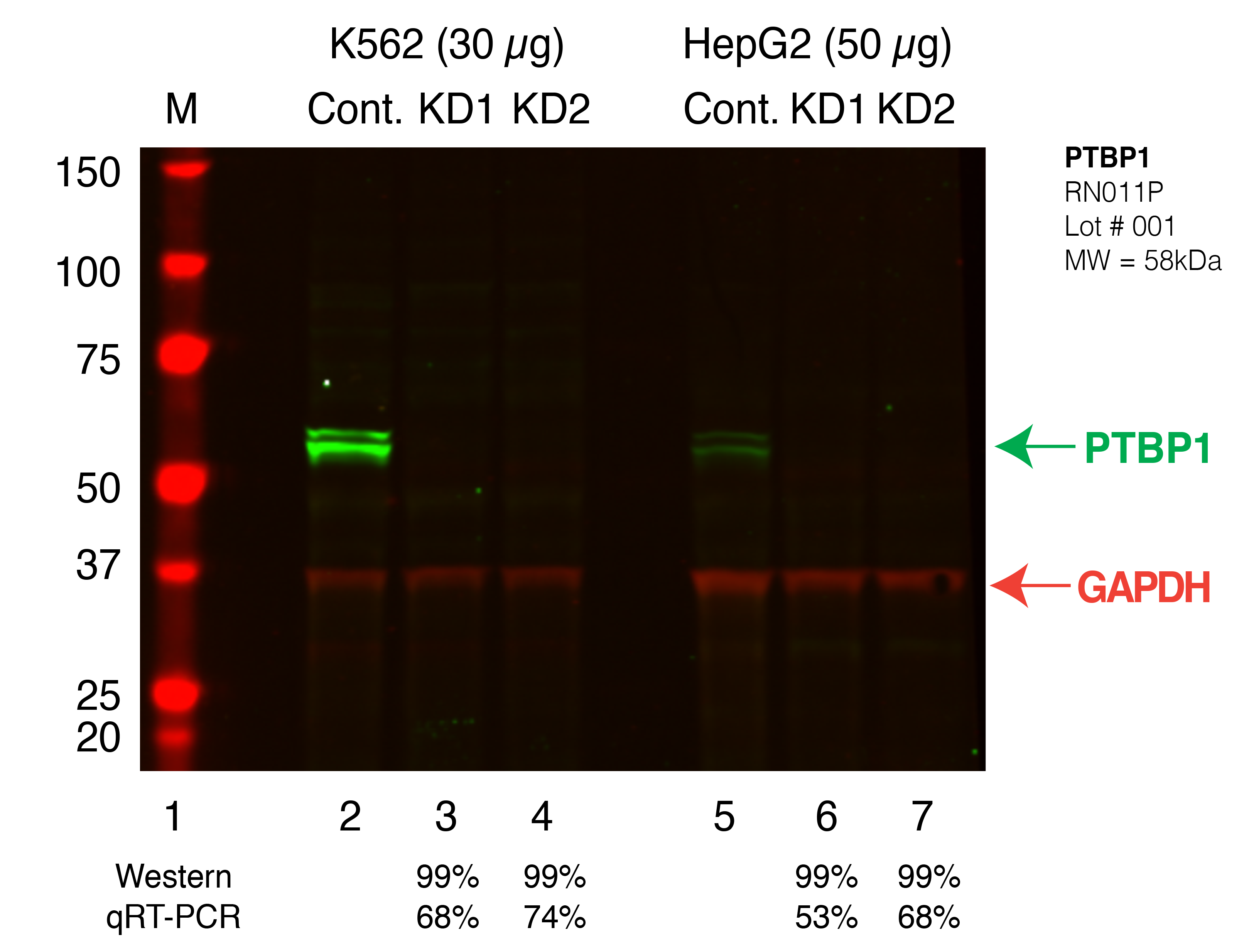
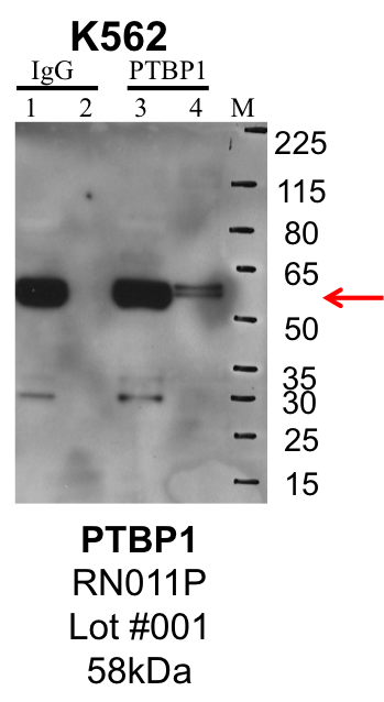
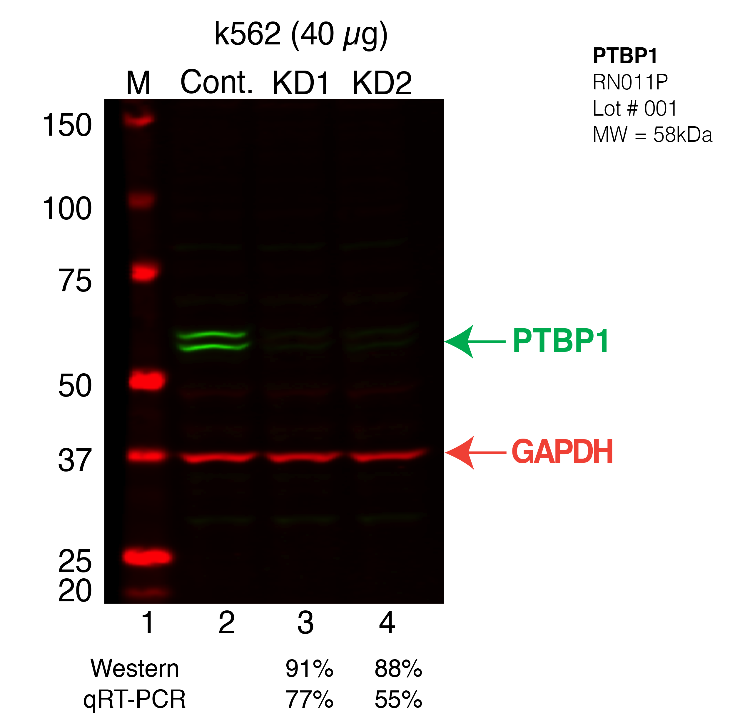
| PTBP1 | Product_ID: RN011P
Lot_ID: 1
Source: MBLI
Target Name: PTBP1-human | 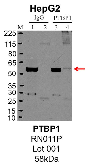
| 
| 
|
| | |
|---|
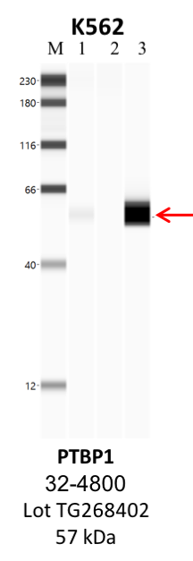
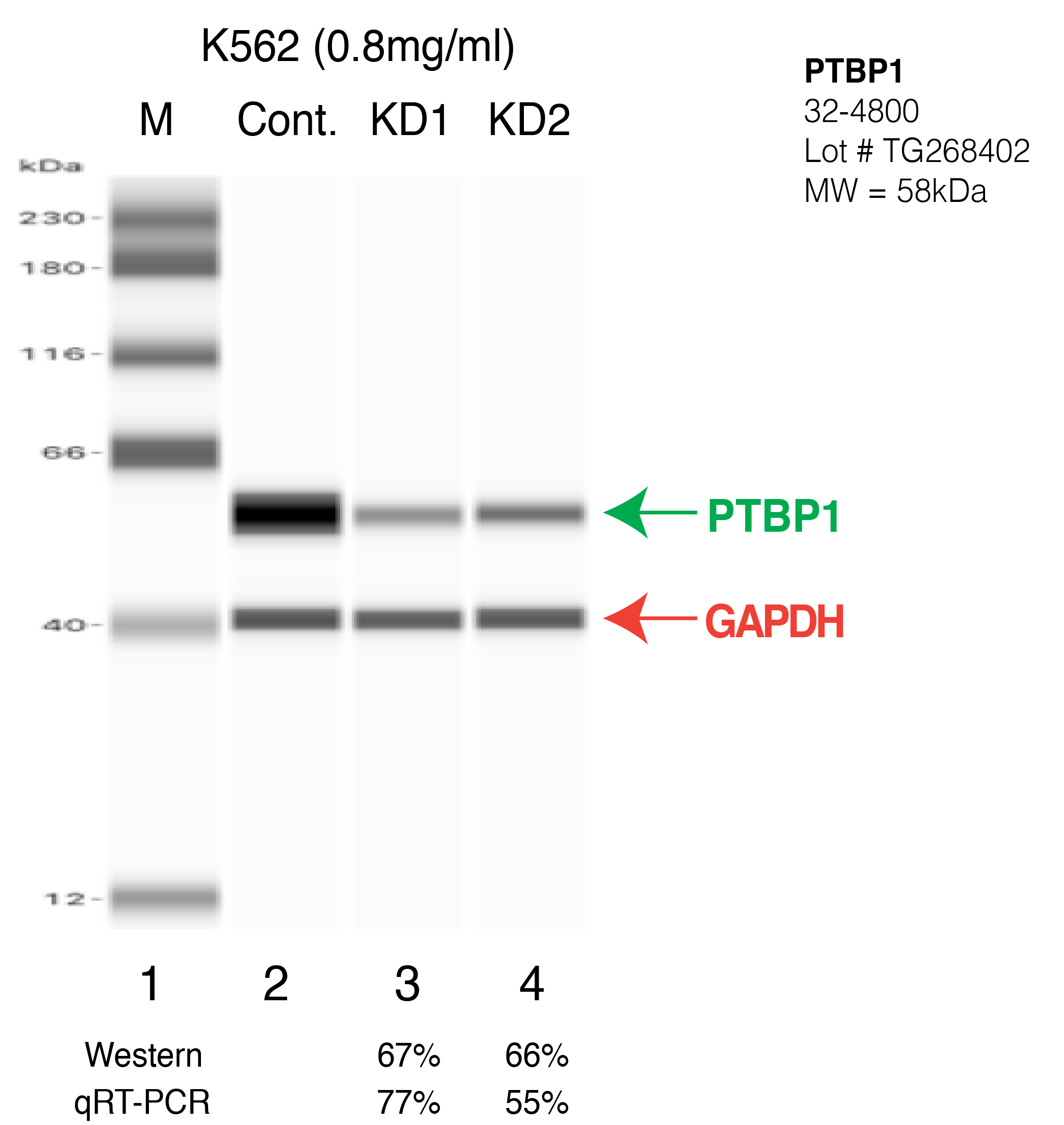
| PTBP1 | Product_ID: 32-4800
Lot_ID: TG268402
Source: Thermo Fisher
Target Name: PTBP1-human | | 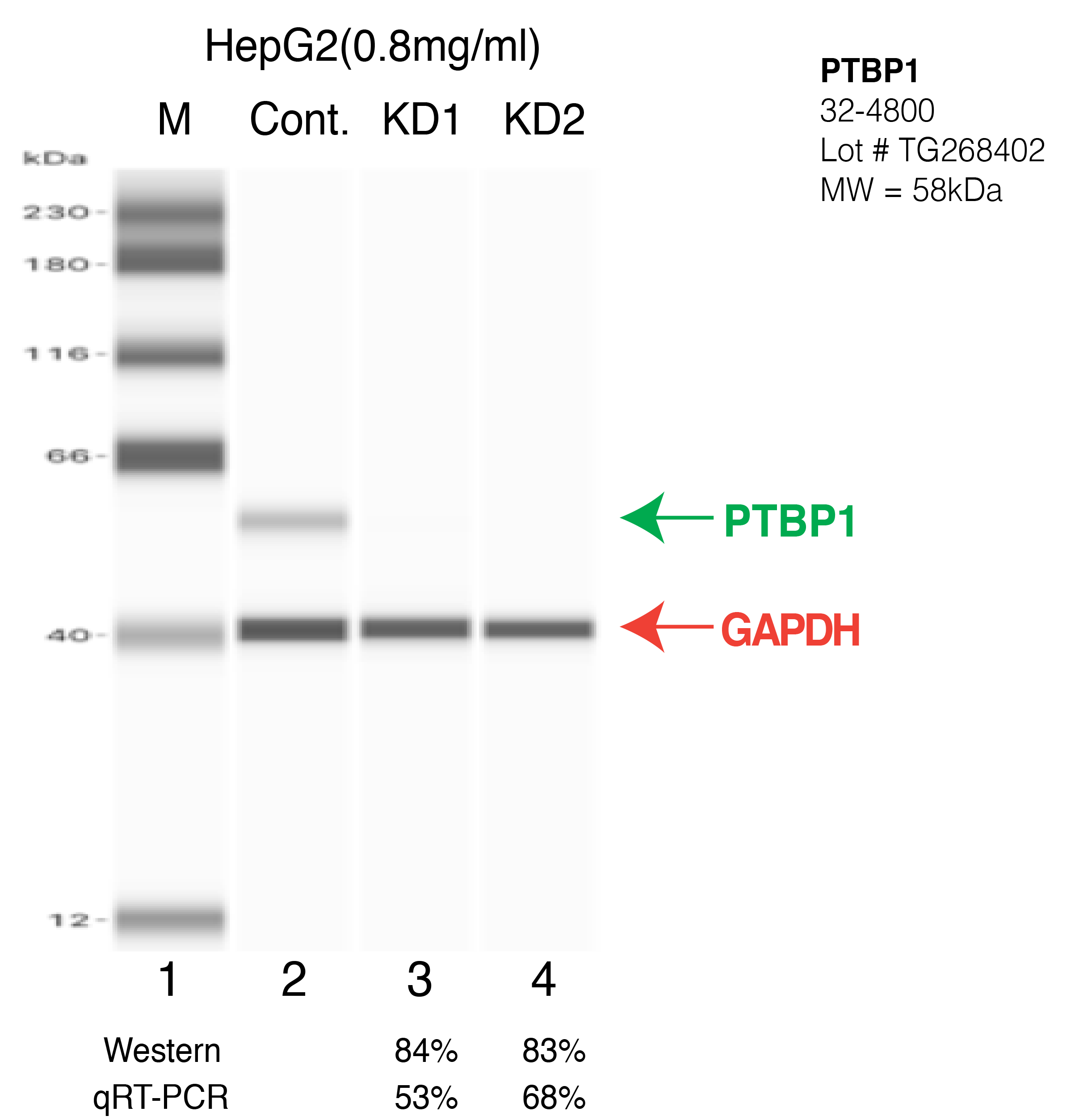
| 
| 
| | |
|---|
| PTBP1 | Product_ID: ARP40421_P050
Lot_ID: QC10301
Source: Aviva
Target Name: PTBP1-human | | | | | | 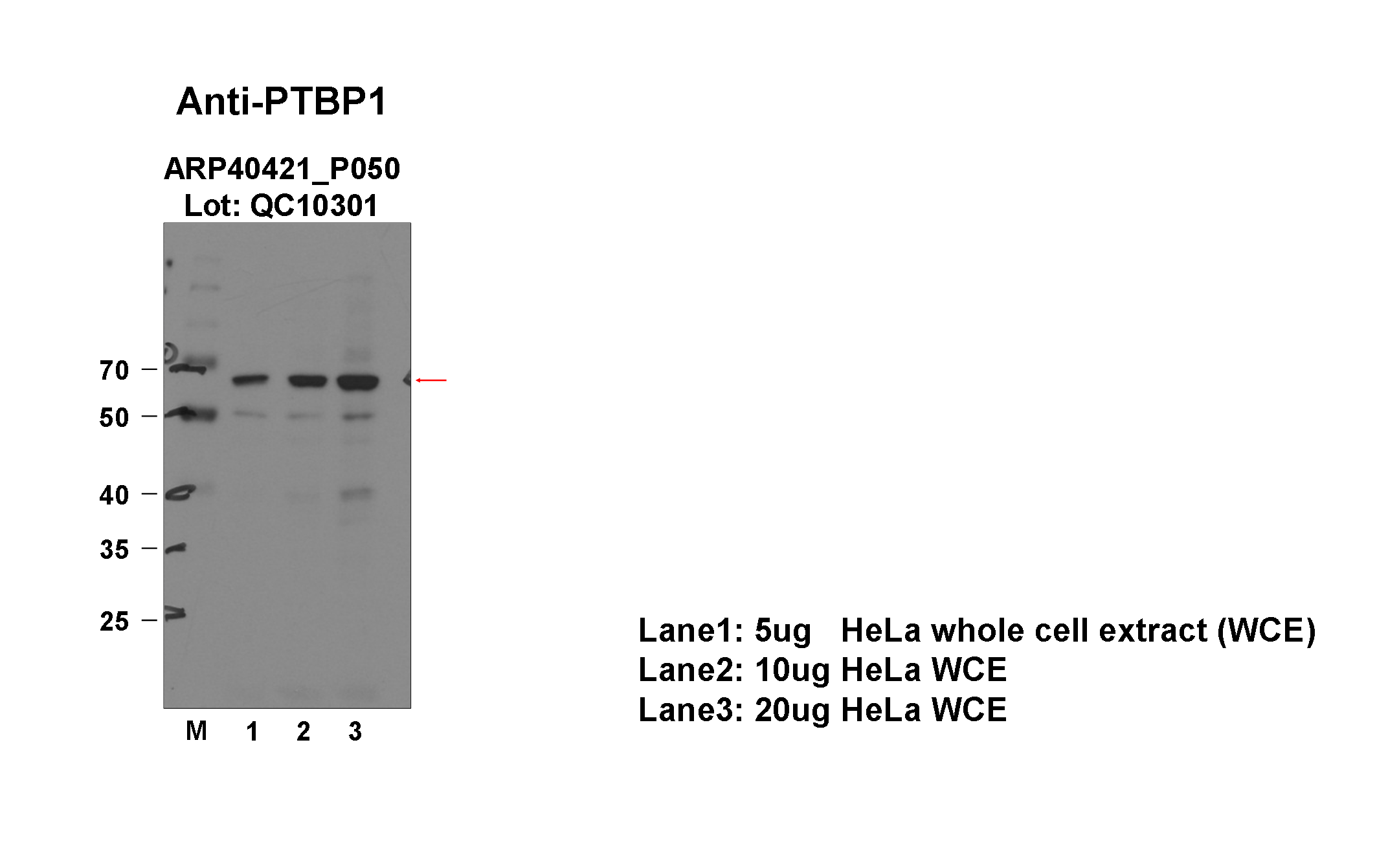
|
|---|
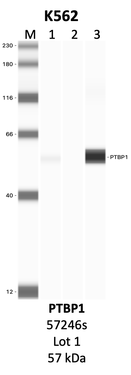
| PTBP1 | Product_ID: 57246S
Lot_ID: 1
Source: Cell Signaling Technology
Target Name: PTBP1-human | | 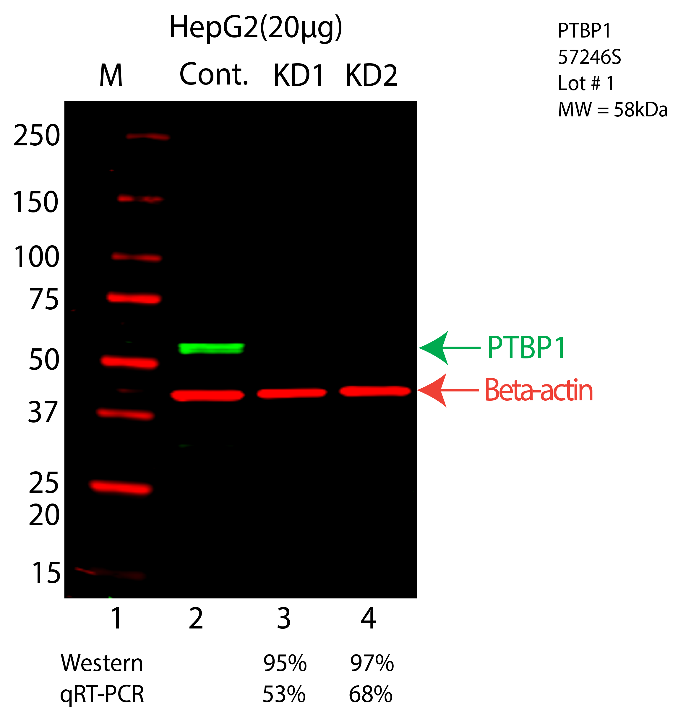
| 
| | | |
|---|
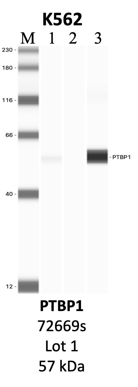
| PTBP1 | Product_ID: 72669S
Lot_ID: 1
Source: Cell Signaling Technology
Target Name: PTBP1-human | | 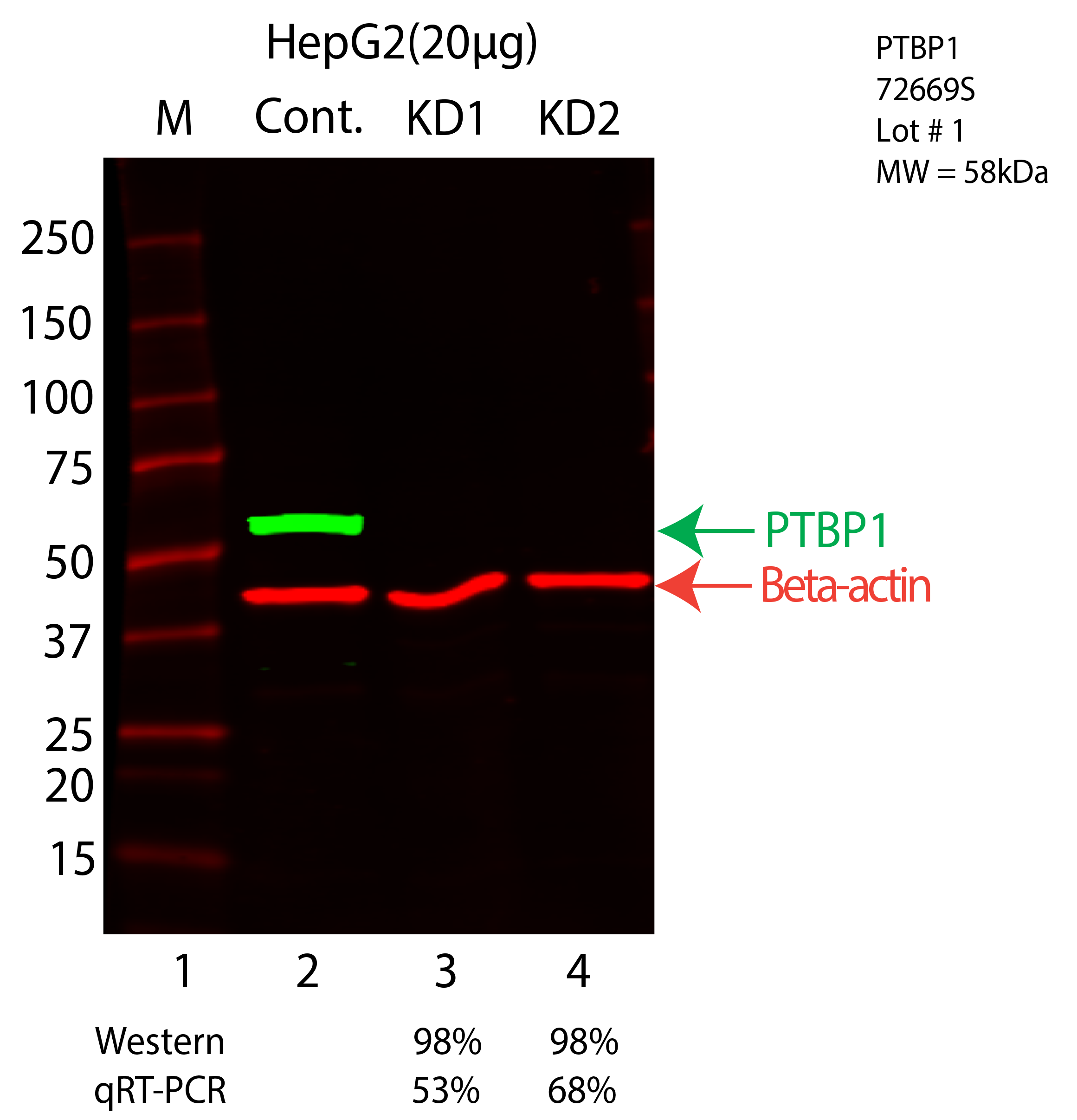
| 
| | | |
|---|