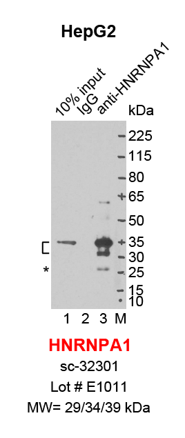
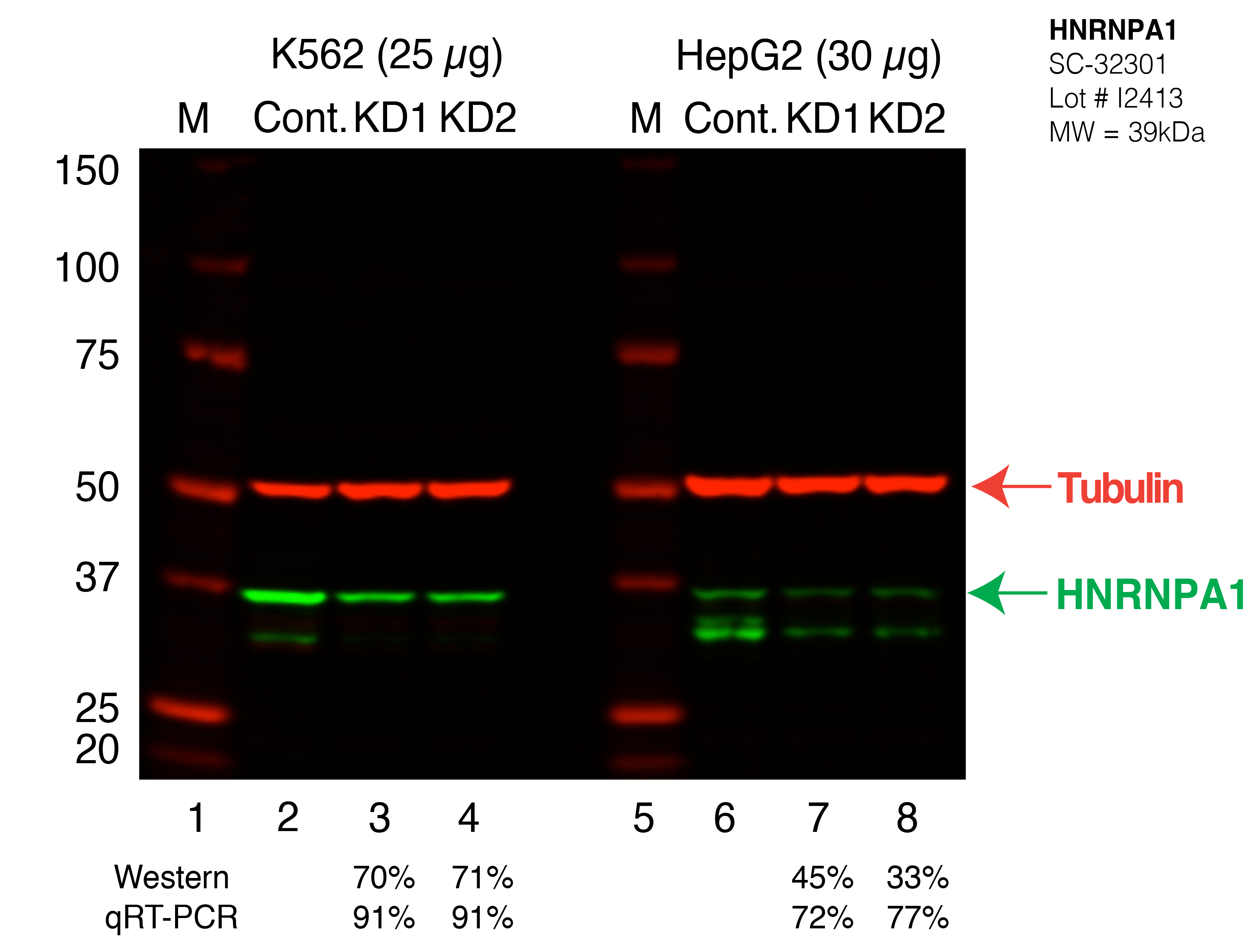
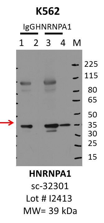
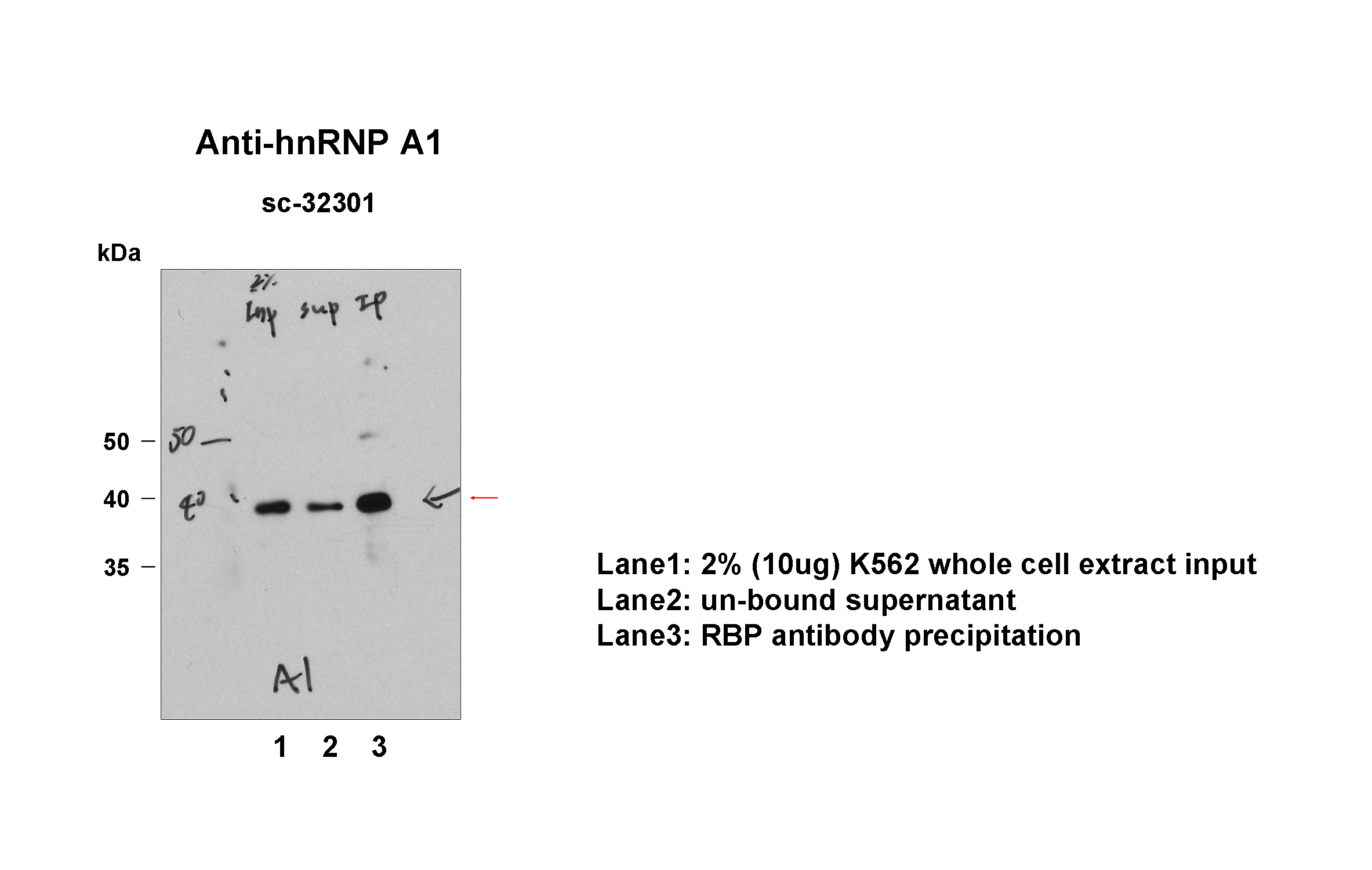
| HNRNPA1 | Product_ID: sc-32301
Lot_ID: I2413
Source: Santa Cruz Biotech
Target Name: HNRNPA1-human | 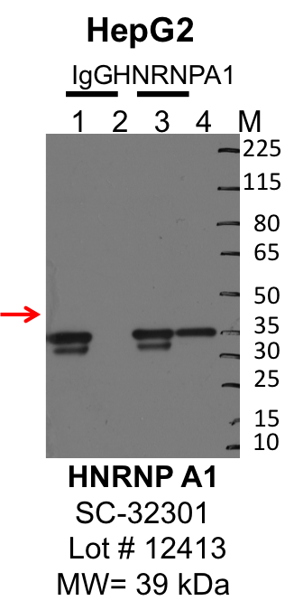
| 
| 
|
| | |
|---|
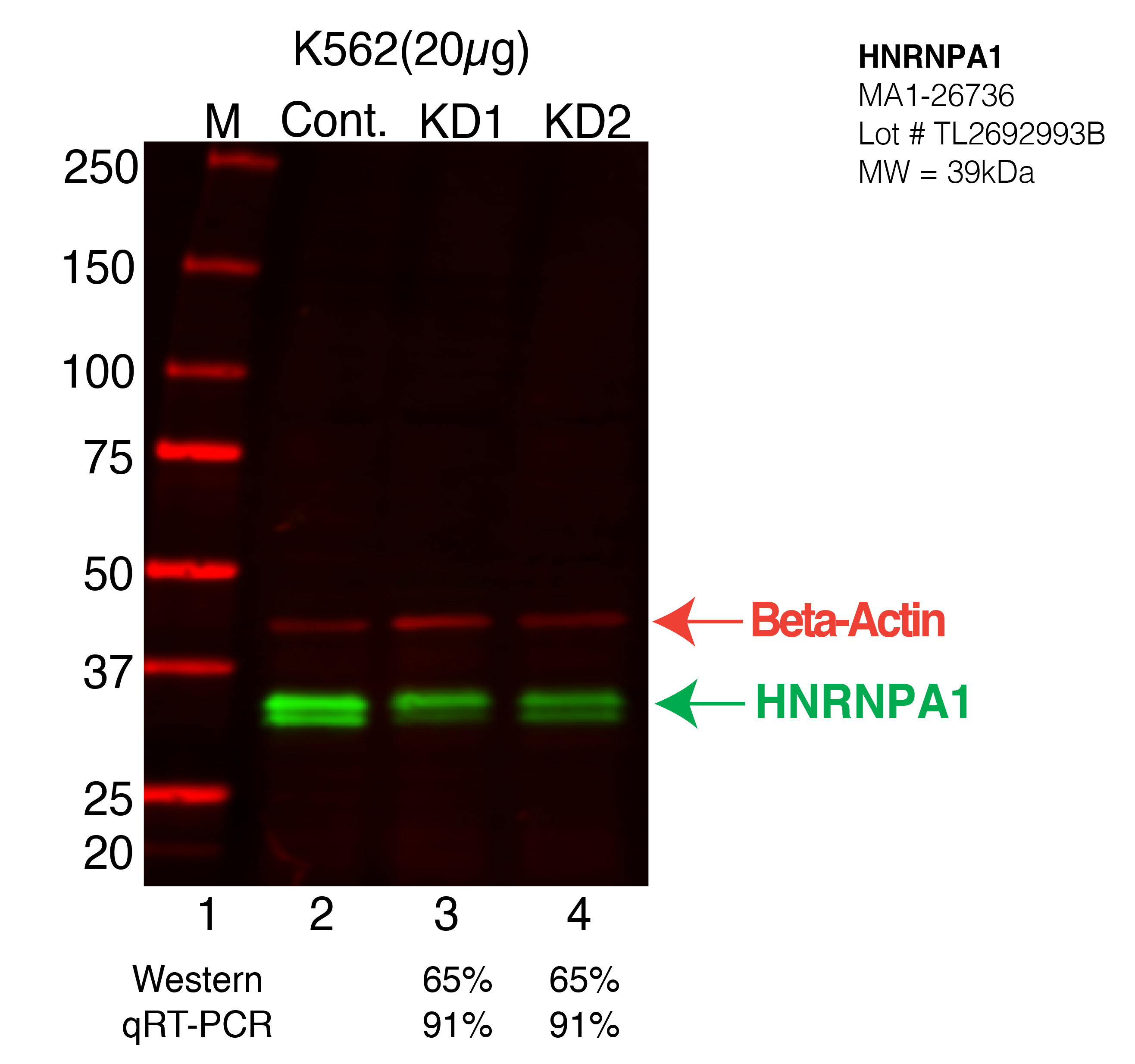
| HNRNPA1 | Product_ID: MA1-26736
Lot_ID: TL2692993B
Source: Thermo Fisher
Target Name: HNRNPA1-human | | | 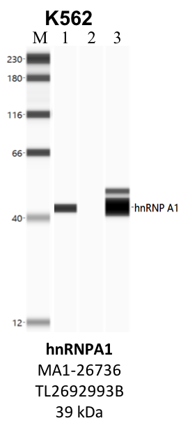
| 
| | |
|---|
| HNRNPA1 | Product_ID: ARP40383_T100
Lot_ID: QC9473-091124
Source: Aviva
Target Name: HNRNPA1-human | | | | | | 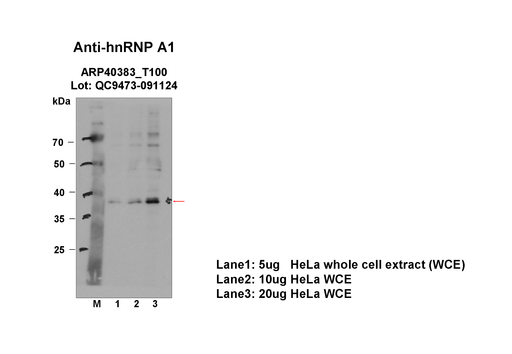
|
|---|
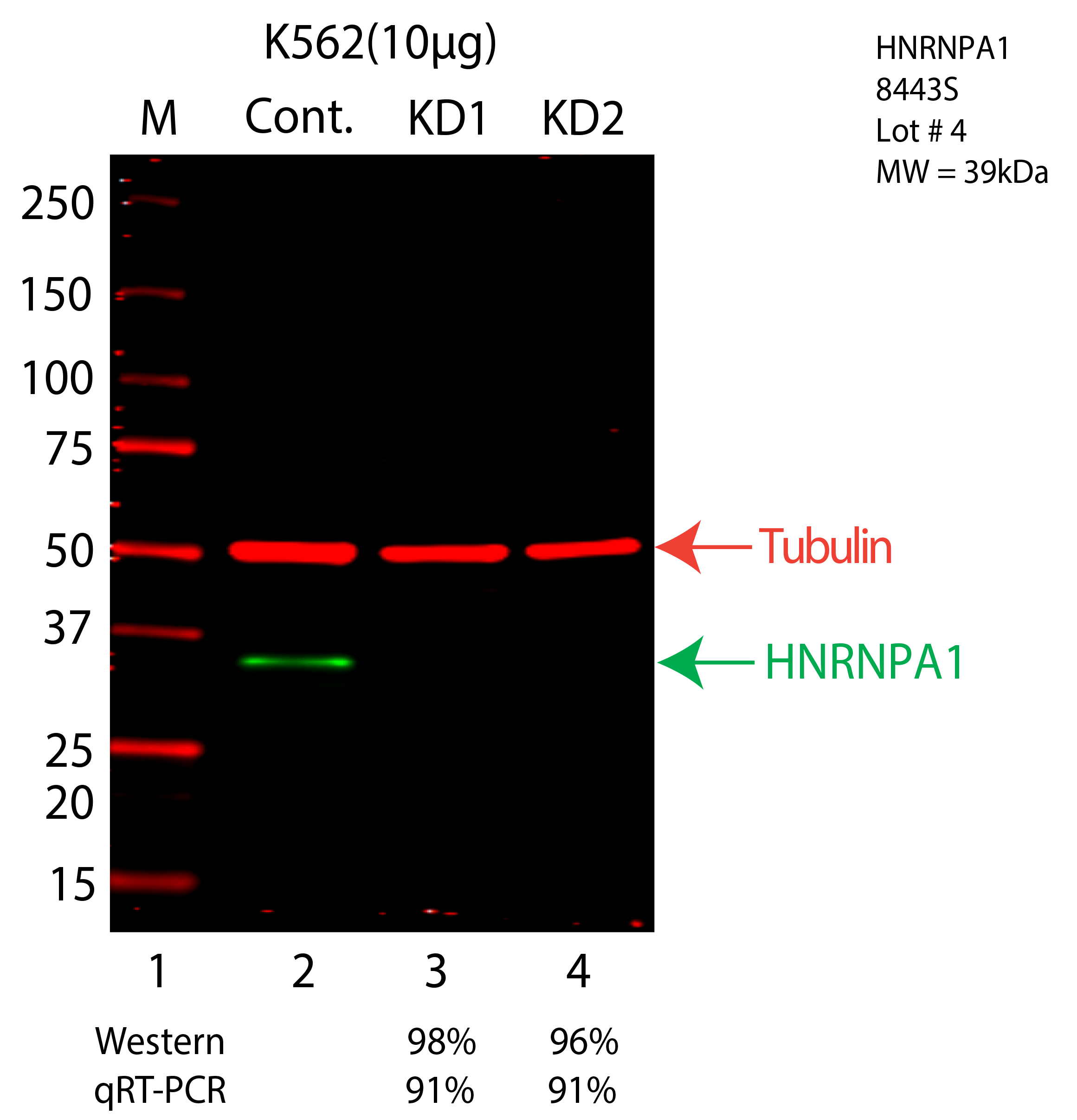
| HNRNPA1 | Product_ID: 8443S
Lot_ID: 4
Source: Cell Signaling Technology
Target Name: HNRNPA1-human | | | 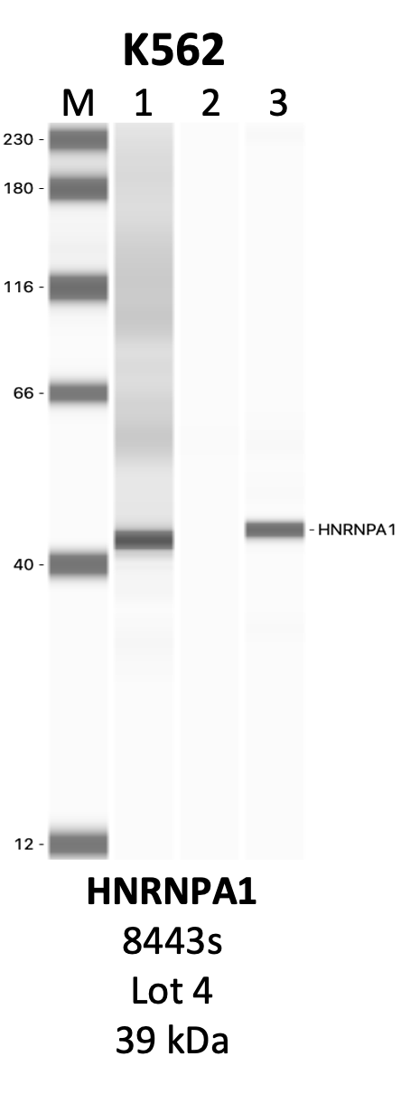
| 
| | |
|---|