| K562 | 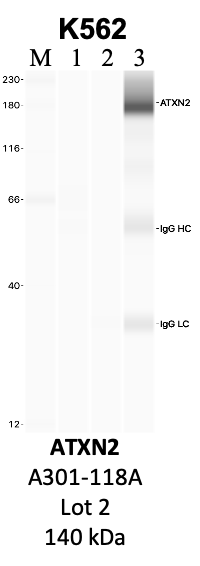
Caption: IP-WB analysis of K562 whole cell lysate using the ATXN2 specific antibody, A301-118A. Lanes 1 and 2 are 2.5% of five million whole cell lysate input and 50% of IP enrichment, respectively, using a normal IgG antibody. Lane 3 is 50% of IP enrichment from five million whole cell lysate using the ATXN2-specific antibody, A301-118A. The same antibody was used to detect protein levels via Western blot. This antibody passes preliminary validation and will be further pursued for secondary validation. *NOTE* Protein sizes are taken from Genecards.org and are only estimates based on sequence. Actual protein size may differ based on protein characteristics and electrophoresis method used. Comments: Immunoprecipitation lane (3) exhibits a large deviation from the expected protein size, which we believe is due to performing the characterization using the Jess Western Blotting system. This system has a documented tendency for some proteins to run differently than with more traditional methods due to their compositions and interactions with the electrophoretic components. The image also closely resembles the example image provided by the vendor. Method: immunoprecipitation Status: Released Lab: Yeo Lab | 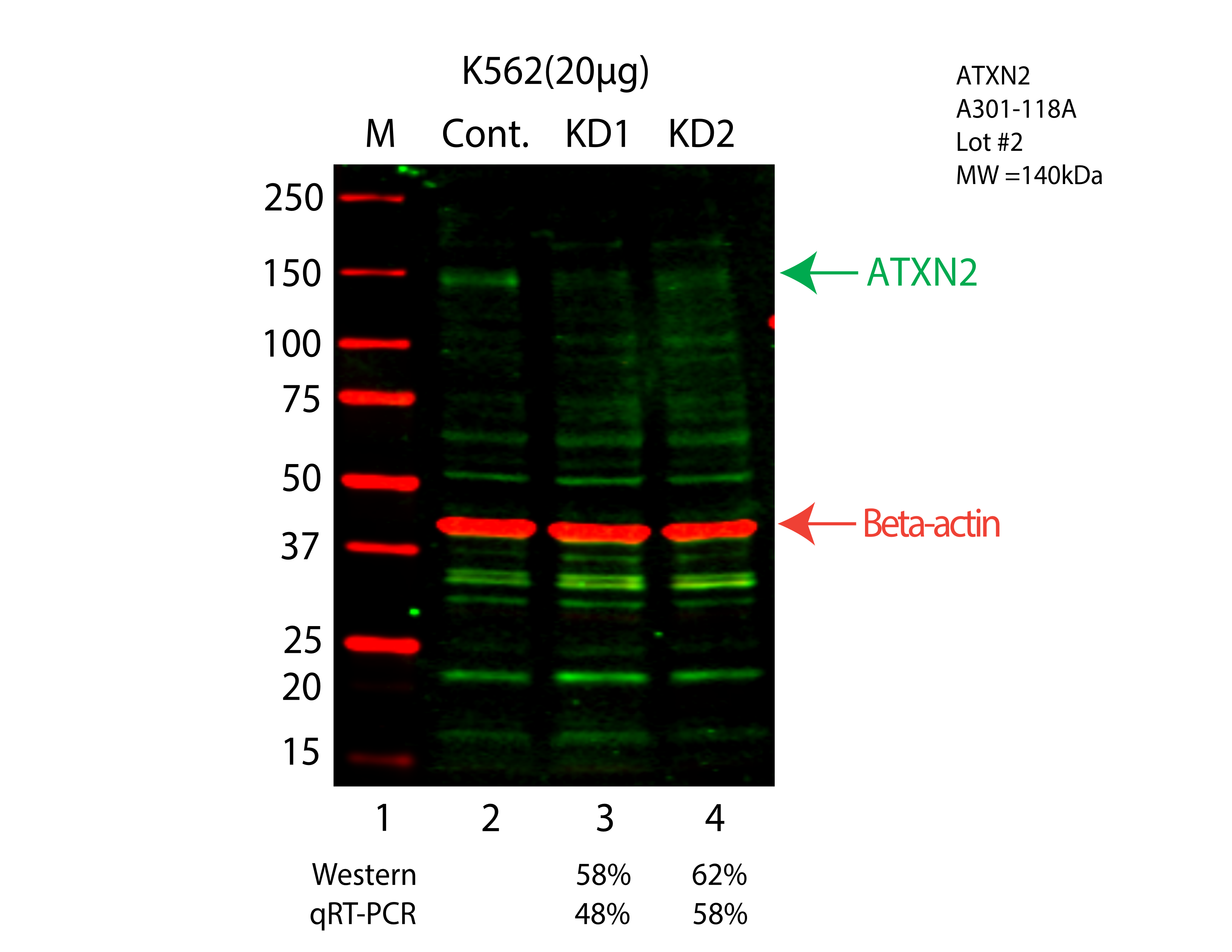
Caption: Western blot following CRISPR against ATXN2 in K562 whole cell lysate using ATXN2 specific antibody. Lane 1 is a ladder, lane 2 is K562 non-targeting control knockdown, lane 3 and 4 are two different CRISPR against ATXN2. ATXN2 protein appears as the green arrow, Beta-actin serves as a control and appears in red arrow. Releated Sample: BGKcLV51-17 Status: Experimented Lab: Graveley Lab |
|---|