| HepG2 | | 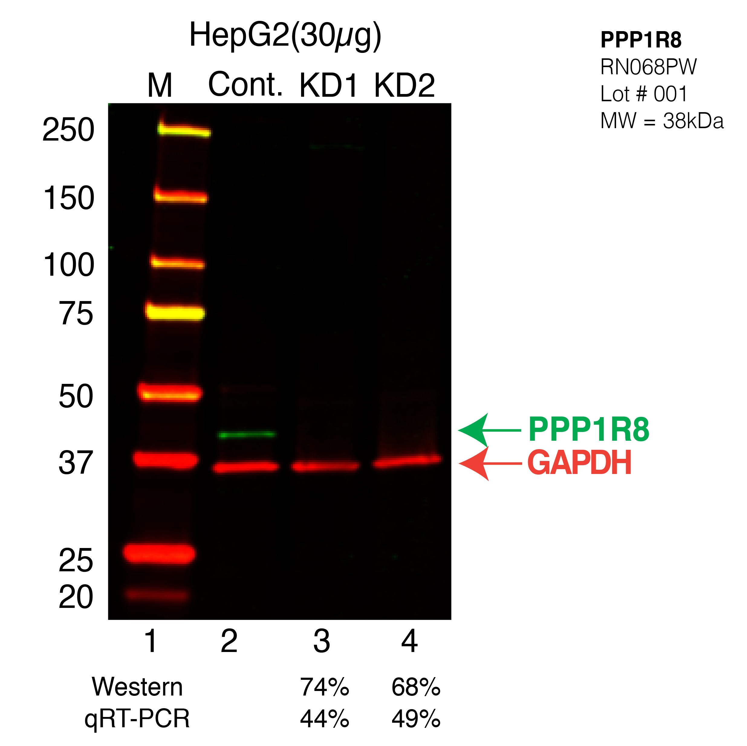
Caption: Western blot following CRISPR against PPP1R8 in HepG2 whole cell lysate using PPP1R8 specific antibody. Lane 1 is a ladder, lane 2 is HepG2 non-targeting control knockdown, lane 3 and 4 are two different CRISPR against PPP1R8. PPP1R8 protein appears as the green band, GAPDH serves as a control and appears in red. Releated Sample: BGHcLV12-63 Status: Released Lab: Graveley Lab |
|---|
| K562 | 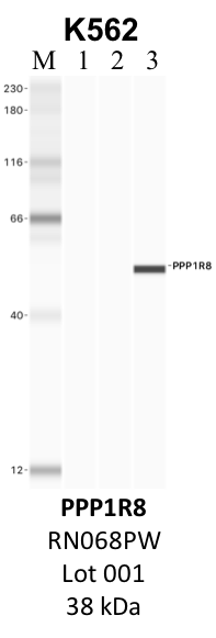
Caption: IP-WB analysis of K562 whole cell lysate using the PPP1R8 specific antibody, RN068PW. Lane 1 is 2.5% of five million whole cell lysate input. Lanes 2 and 3 are 50% of IP enrichment from five million whole cell lysate using normal IgG antibody and the PPP1R8-specific antibody, RN068PW. The same antibody was used to detect protein levels via Western blot. This antibody passes preliminary validation and will be further pursued for secondary validation. *NOTE* Protein sizes are taken from Genecards.org and are only estimates based on sequence. Actual protein size may differ based on protein characteristics and electrophoresis method used. Comments: Immunoprecipitation lane (3) exhibits a large deviation from the expected protein size, which we believe is due to performing the characterization using the Jess Western Blotting system. This system has a documented tendency for some proteins to run differently than with more traditional methods due to their compositions and interactions with the electrophoretic components. Method: immunoprecipitation Status: NotSatisfied Lab: Yeo Lab | 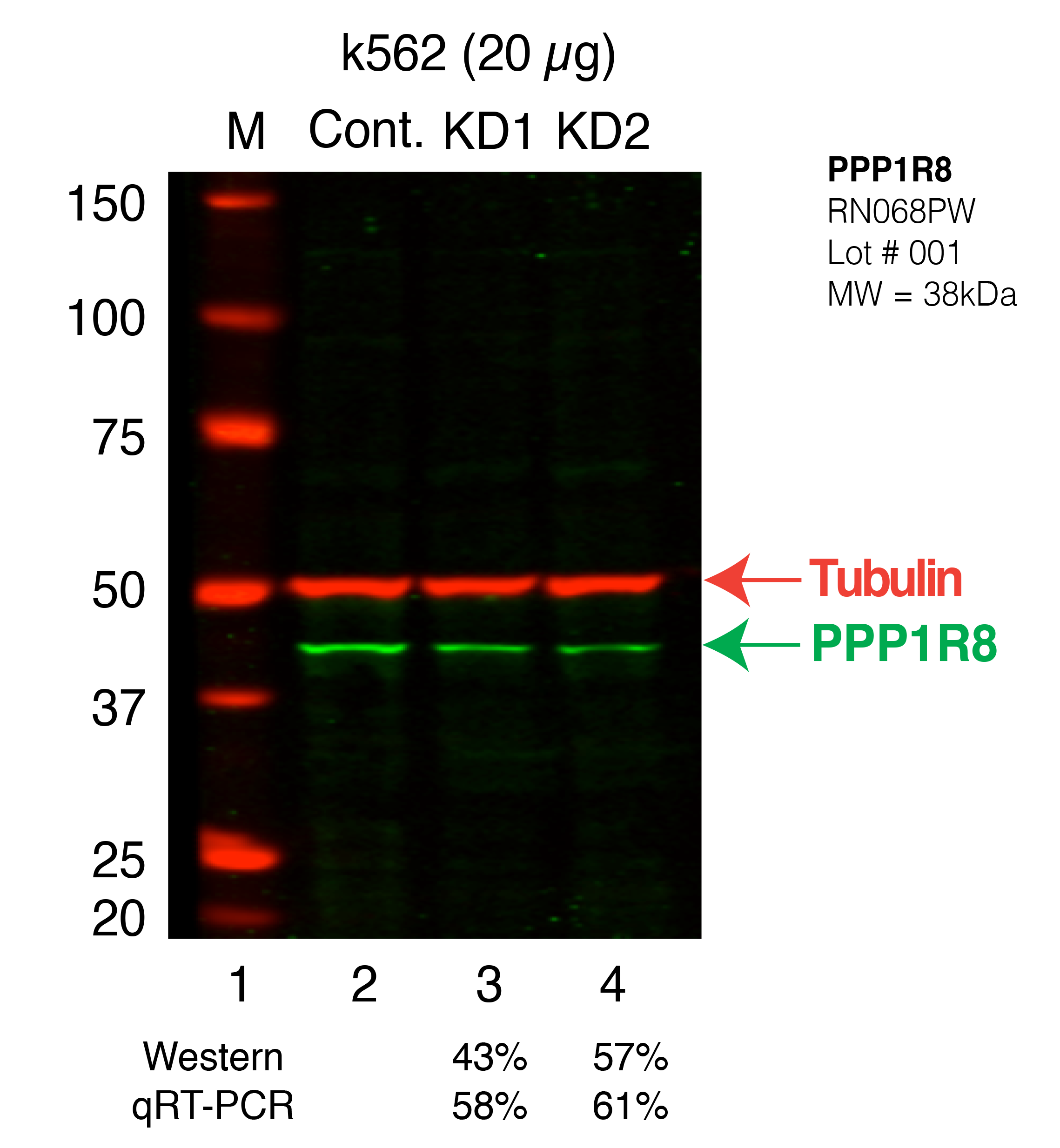
Caption: Western blot following shRNA against PPP1R8 in K562 whole cell lysate using PPP1R8 specific antibody. Lane 1 is a ladder, lane 2 is K562 non-targeting control knockdown, lane 3 and 4 are two different shRNAs against PPP1R8. PPP1R8 protein appears as the green band, Tubulin serves as a control and appears in red. Releated Sample: BGKLV28-41 Status: Released Lab: Graveley Lab |
|---|