| HepG2 | 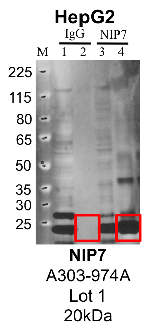
Caption: Representative image of immunoprecipitation performed on whole cell extracts from the HepG2 cell line using the NIP7-specific antibody A303-974A. Lane 1: Input from IP using control IgG. Lane 2: Immunoprecipitated material using control IgG. Lane 3: Input from IP using NIP7 antibody. Lane 4: Immunoprecipitated material using NIP7 antibody. Outlined regions were excised from gel and subjected to analysis by mass spectrometry. Target molecular weight: 20.46 kDa. Method: immunoprecipitation Releated Sample: eCLIP:703 Status: Released Lab: Yeo Lab 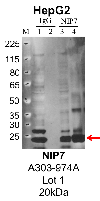
Caption: IP-Western Blot analysis of HepG2 whole cell lysate using NIP7 specific antibody. Lane 1 is 1% of twenty million whole cell lysate input and lane 2 is 25% of IP enrichment using rabbit normal IgG (lanes under 'IgG'). Lane 3 is 1% of twenty million whole cell lysate input and lane 4 is 10% IP enrichment using rabbit polyclonal anti-NIP7 antibody (lanes under 'NIP7'). Method: immunoprecipitation Releated Sample: eCLIP:703 Status: Released Lab: Yeo Lab | 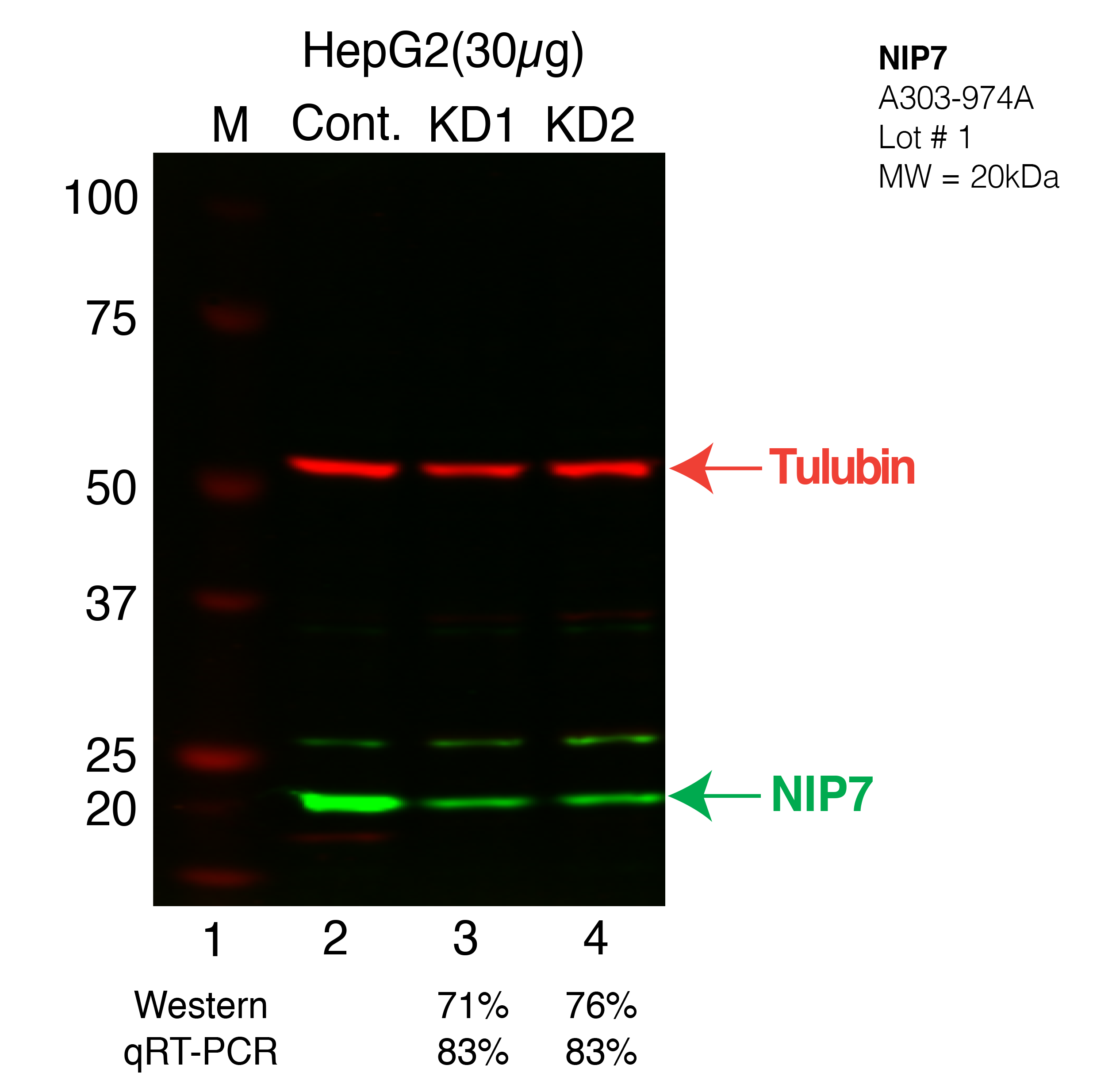
Caption: Western blot following CRISPR against NIP7 in HepG2 whole cell lysate using NIP7 specific antibody. Lane 1 is a ladder, lane 2 is HepG2 non-targeting control knockdown, lane 3 and 4 are two different CRISPR against NIP7.NIP7 protein appears as the green band, Tubulin serves as a control and appears in red. Releated Sample: BGHcLV09-5 Status: Released Lab: Graveley Lab 
Caption: IP followed by mass spectrometry. Protein was immunoprecipitated from HepG2 whole cell lysates using the antibody A303-974A, loaded on a 4-12% NuPAGE Bis-Tris gel, and separated via electrophoresis. Using a reference western blot done in parallel, gel pieces corresponding to the sections indicated were excised and submitted for analysis by the UCSD Biomolecular and Proteomics Mass Spectrometry Facility. Status: Released Lab: Yeo Lab |
|---|
| K562 | | 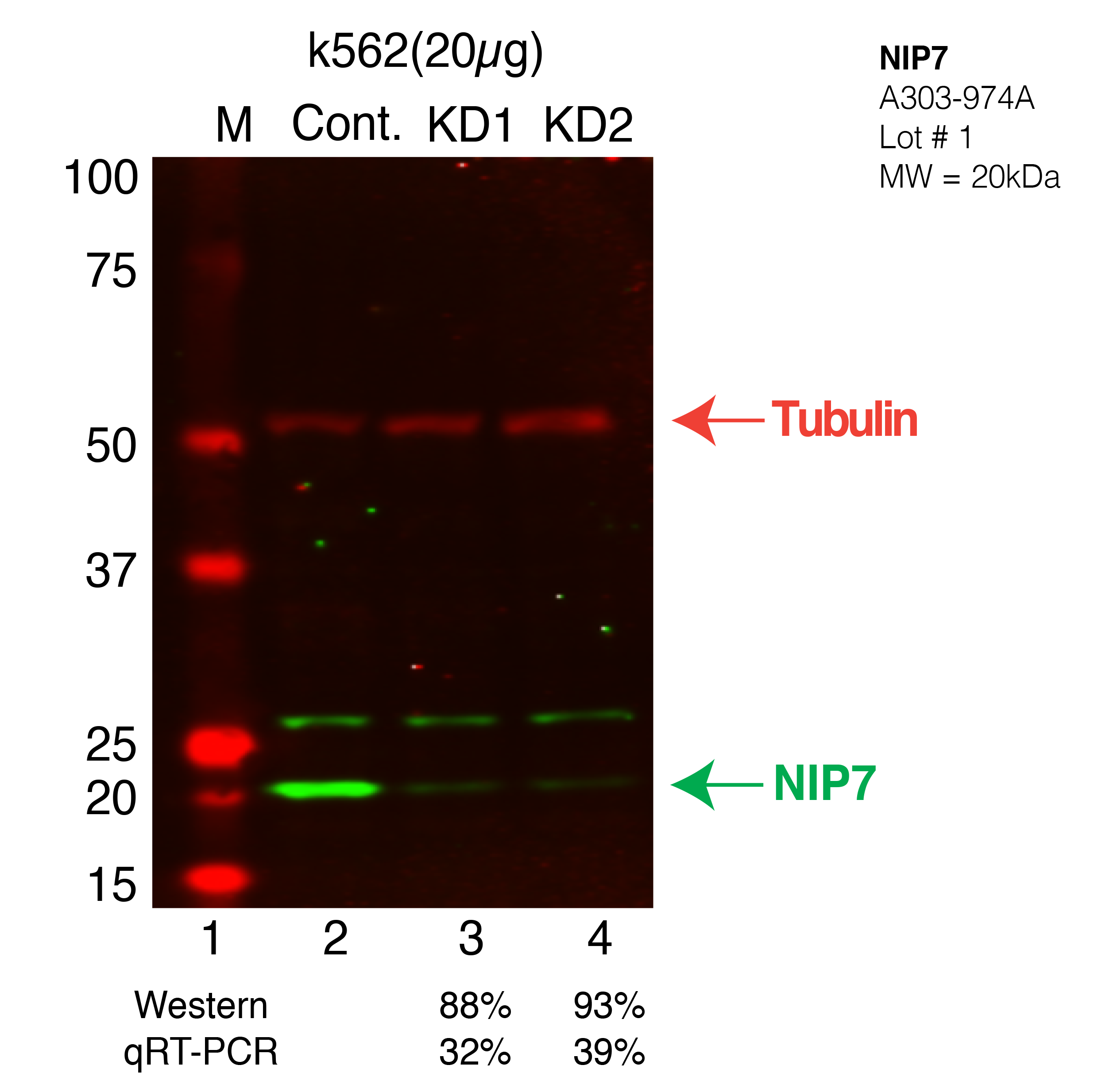
Caption: Western blot following CRISPR against NIP7 in K562 whole cell lysate using NIP7 specific antibody. Lane 1 is a ladder, lane 2 is K562 non-targeting control knockdown, lane 3 and 4 are two different CRISPR against NIP7.NIP7 protein appears as the green band, Tubulin serves as a control and appears in red. Releated Sample: BGKcLV11-67 Status: Released Lab: Graveley Lab |
|---|