| HepG2 | | 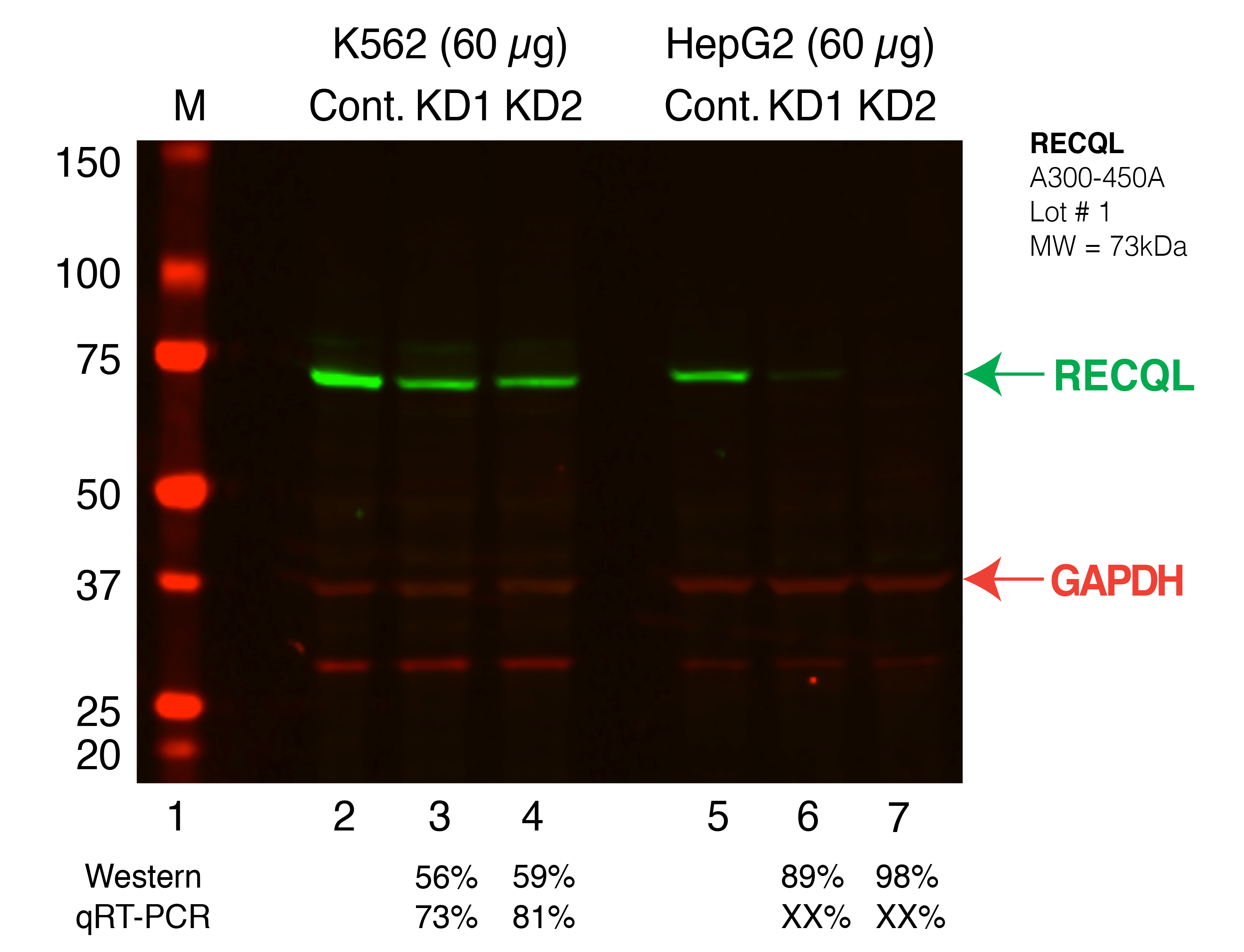
Caption: Western blot following shRNA against RECQL in K562 and HepG2 whole cell lysate using RECQL specific antibody. Lane 1 is a ladder, lane 2 is K562 non-targeting control knockdown, lane 3 and 4 are two different shRNAs against RECQL. Lanes 5-7 follow the same pattern, but in HepG2. RECQL protein appears as the green band, GAPDH serves as a control and appears in red. Releated Sample: BGKLV08-49 Status: Released Lab: Graveley Lab |
|---|
| K562 | |
Caption: Western blot following shRNA against RECQL in K562 and HepG2 whole cell lysate using RECQL specific antibody. Lane 1 is a ladder, lane 2 is K562 non-targeting control knockdown, lane 3 and 4 are two different shRNAs against RECQL. Lanes 5-7 follow the same pattern, but in HepG2. RECQL protein appears as the green band, GAPDH serves as a control and appears in red. Releated Sample: BGKLV08-49 Status: Released Lab: Graveley Lab |
|---|