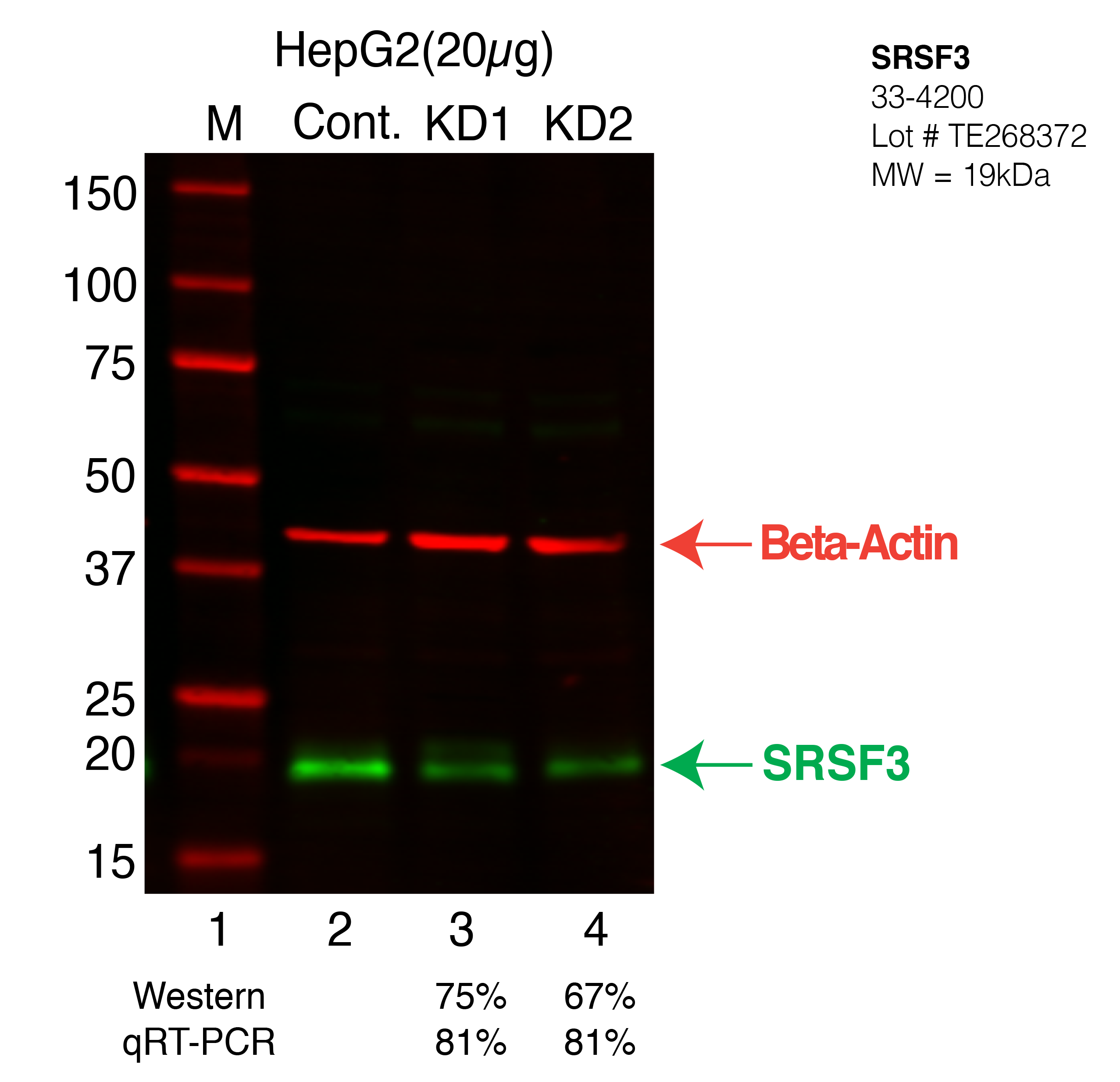
Caption: Western blot following shRNA against SRSF3 in HepG2 whole cell lysate using SRSF3 specific antibody. Lane 1 is a ladder, lane 2 is HepG2 non-targeting control knockdown, lane 3 and 4 are two different shRNA against SRSF3. SRSF3 protein appears as the green arrow, Beta-actin serves as a control and appears in red arrow. Releated Sample: BGHLV35-49 Status: Released Lab: Graveley Lab |