Home
NEWS
Summary
Data Files
Search
FAQ
Admin Login
Top
Antibodies of SUCLG1
2
antibodies are included.
Released
NotSatisfied
Submitted
NotReviewed
OnHold
RequestToSubmit
Experimented
RBP
Antibody Info
Primary-HepG2
Secondary-HepG2
Primary-K562
Secondary-K562
Primary-UBERON
Primary-Hela
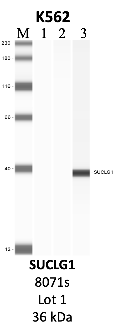
SUCLG1
Product_ID:
GTX109215
Lot_ID: 40023
Source: GeneTex
Target Name: SUCLG1-human
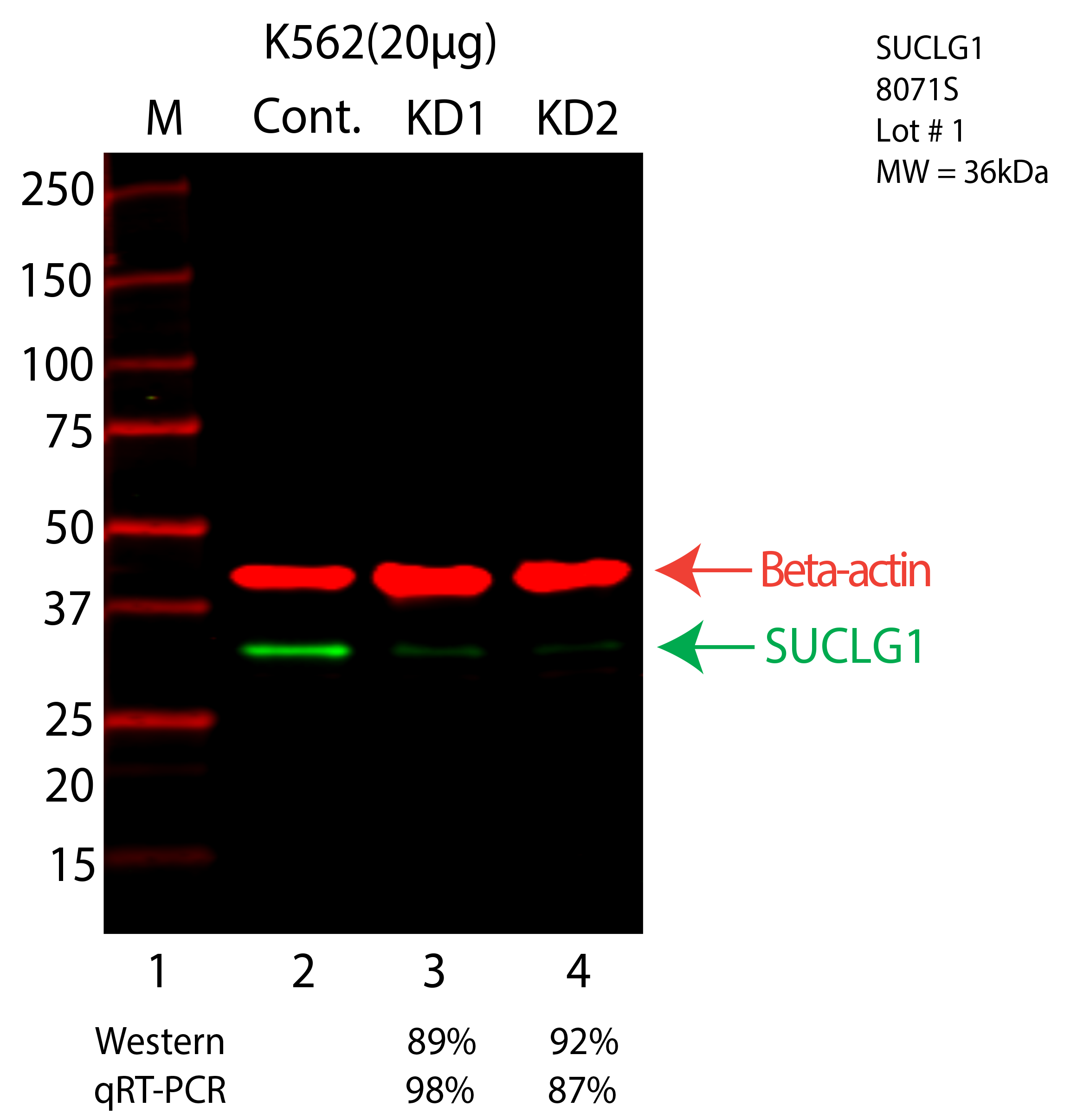
SUCLG1
Product_ID:
8071S
Lot_ID: 1
Source: Cell Signaling Technology
Target Name: SUCLG1-human
×