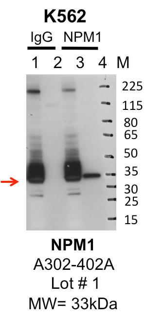
| NPM1 | Product_ID: A302-402A
Lot_ID: 1
Source: Bethyl Labs
Target Name: NPM1-human | | 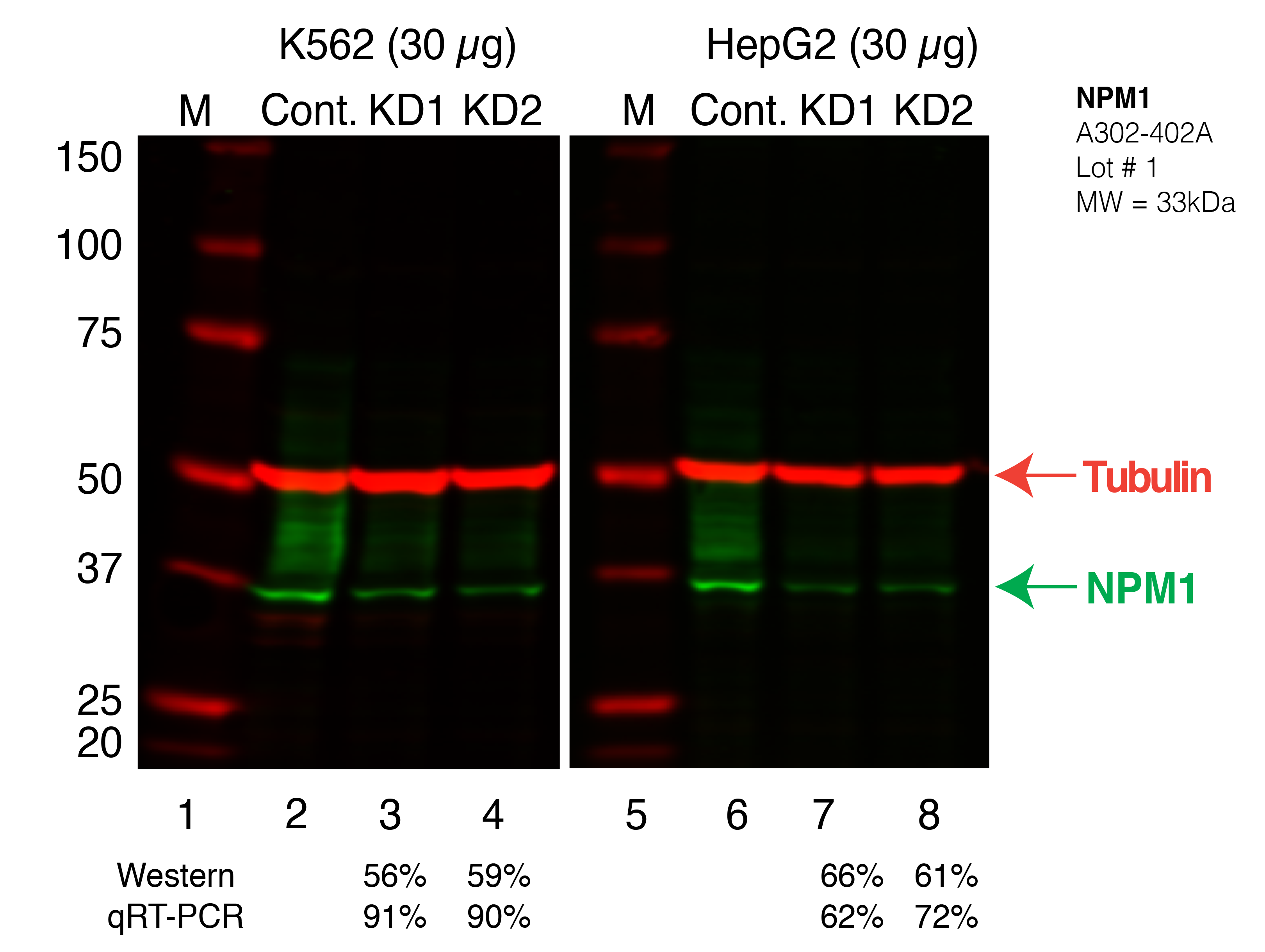
| 
|
| | |
|---|
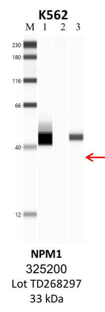
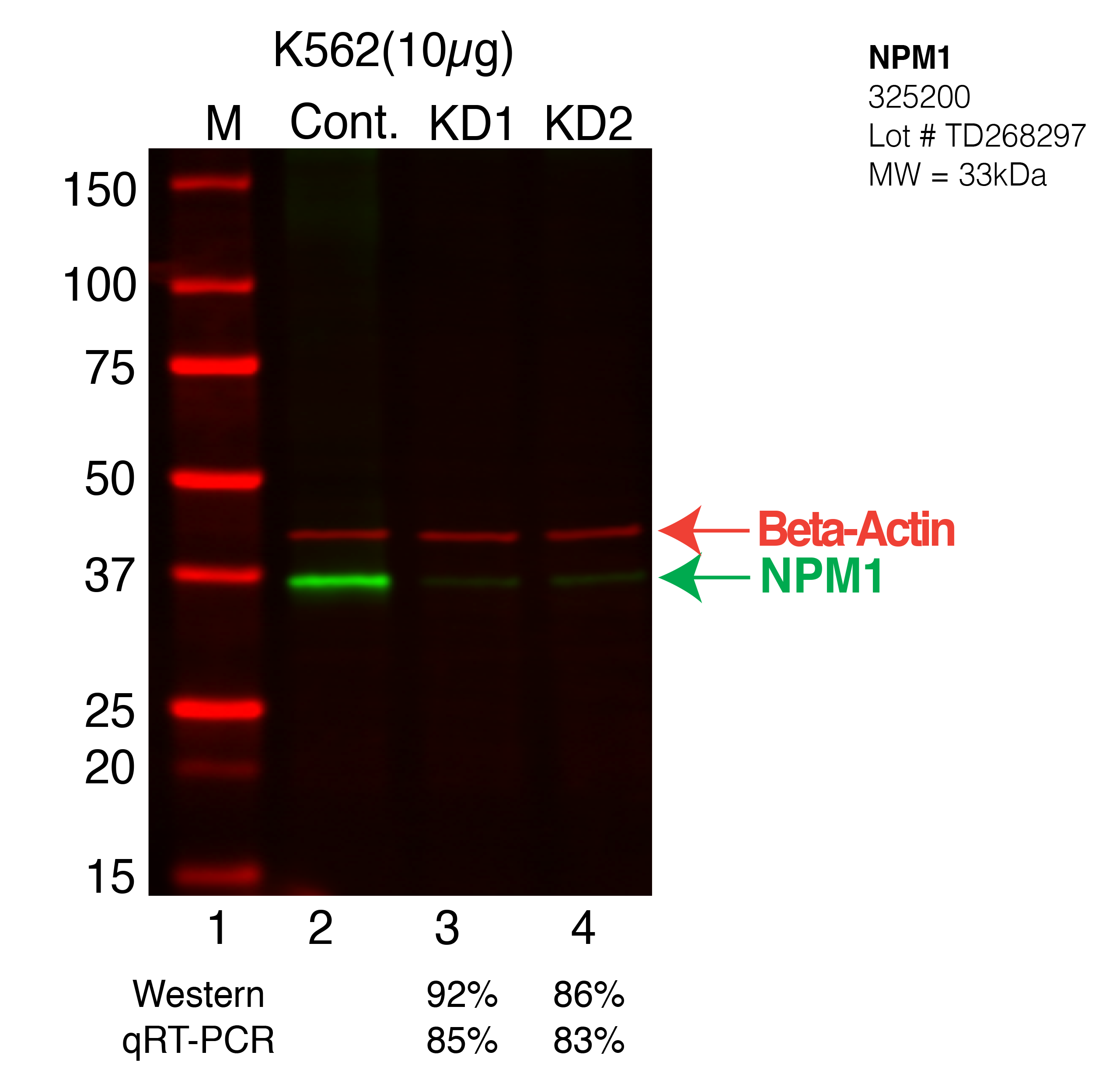
| NPM1 | Product_ID: 325200
Lot_ID: TD268297
Source: Thermo Fisher
Target Name: NPM1-human | | 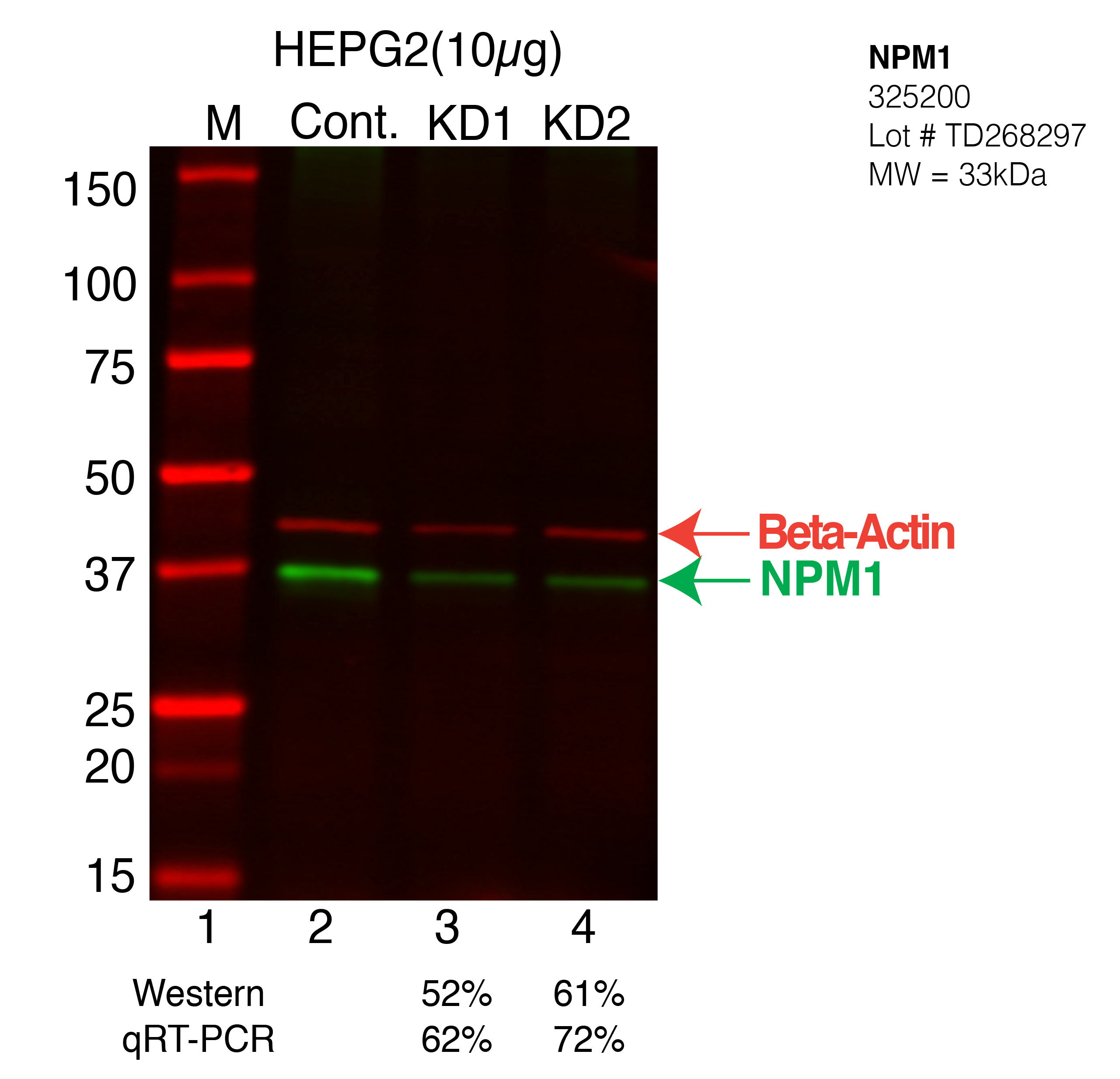
| 
| 
| | |
|---|
| NPM1 | Product_ID: 200143
Lot_ID: AD0811
Source: Zen Bioscience
Target Name: NPM1-human | 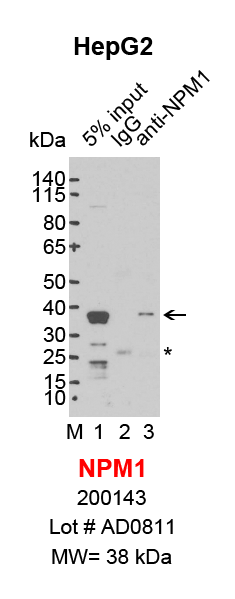
| | | | | |
|---|