Home
NEWS
Summary
Data Files
Search
FAQ
Admin Login
Top
Antibodies of MBNL1
2
antibodies are included.
Released
NotSatisfied
Submitted
NotReviewed
OnHold
RequestToSubmit
RBP
Antibody Info
Primary-HepG2
Secondary-HepG2
Primary-K562
Secondary-K562
Primary-UBERON
Primary-Hela
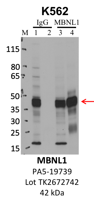
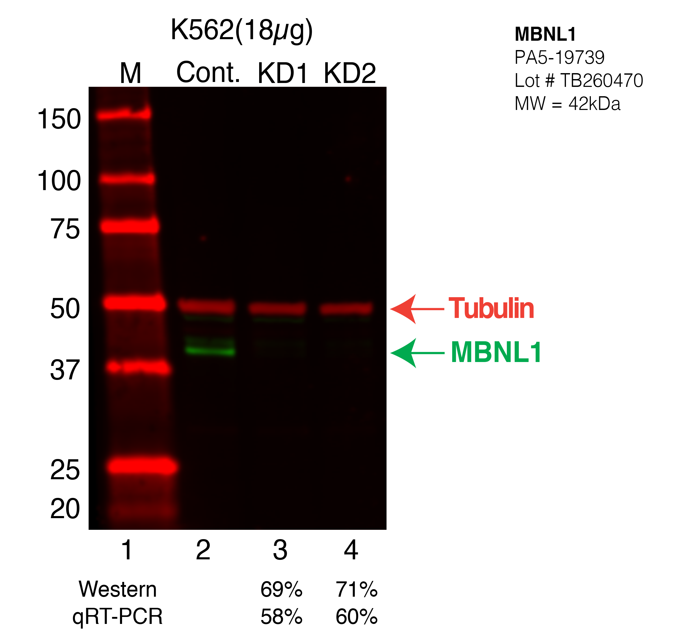
MBNL1
Product_ID:
PA5-19739
Lot_ID: TK2672742
Source: Thermo Fisher
Target Name: MBNL1-human
MBNL1
Product_ID:
GTX112624
Lot_ID: 40562
Source: GeneTex
Target Name: MBNL1-human
×