Home
NEWS
Summary
Data Files
Search
FAQ
Admin Login
Top
Antibodies of FIP1L1
2
antibodies are included.
Released
NotSatisfied
Submitted
NotReviewed
OnHold
RequestToSubmit
Experimented
RBP
Antibody Info
Primary-HepG2
Secondary-HepG2
Primary-K562
Secondary-K562
Primary-UBERON
Primary-Hela
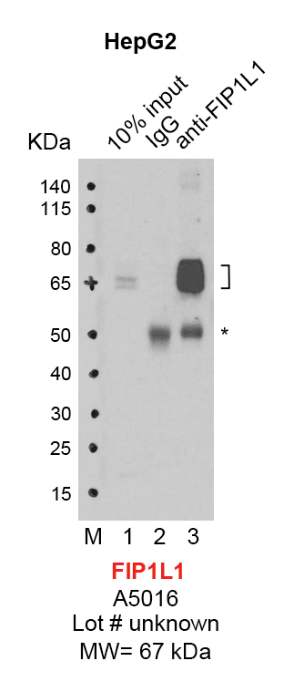
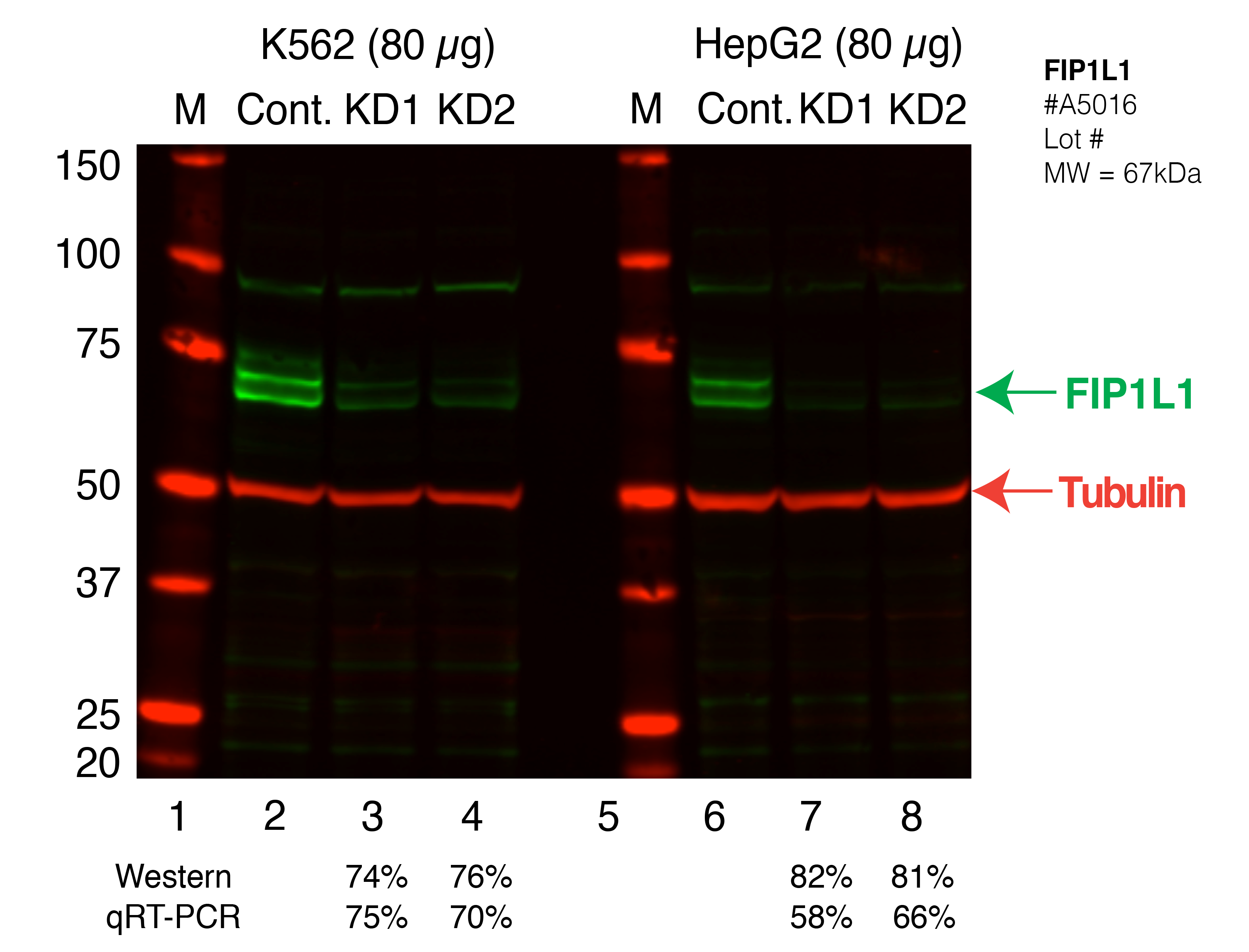
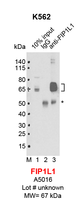
FIP1L1
Product_ID:
A301-461A
Lot_ID: 1
Source: Bethyl Labs
Target Name: FIP1L1-human
FIP1L1
Product_ID:
A5016
Lot_ID: unknown
Source: ABclonal
Target Name: FIP1L1-human
×