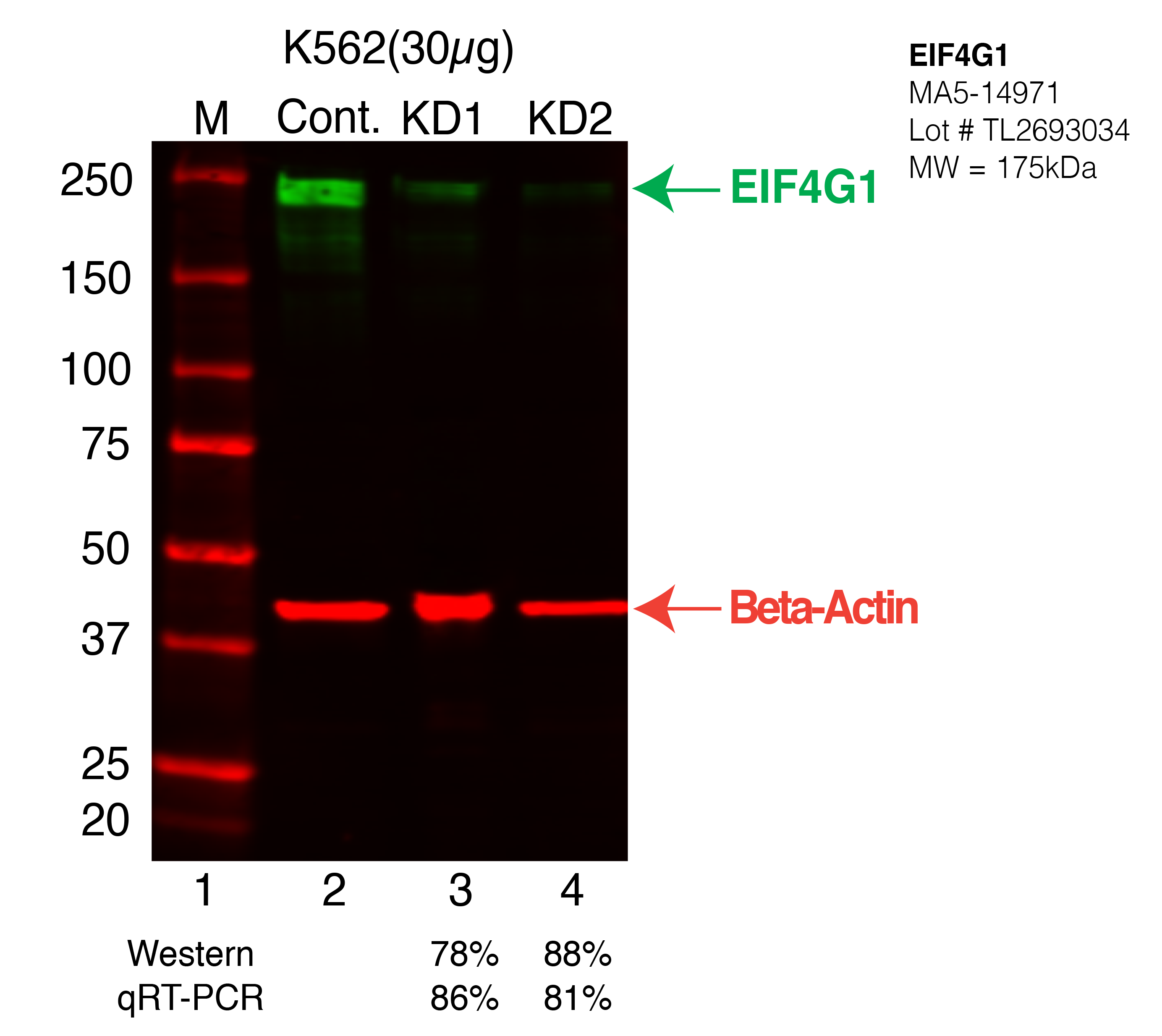
| EIF4G1 | Product_ID: MA5-14971
Lot_ID: TL2693034
Source: Thermo Fisher
Target Name: EIF4G1-human | | | 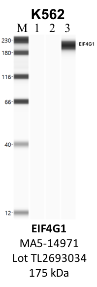
| 
| | |
|---|
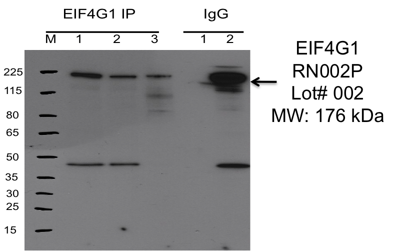
| EIF4G1 | Product_ID: RN002P
Lot_ID: 2
Source: MBLI
Target Name: EIF4G1-human | | 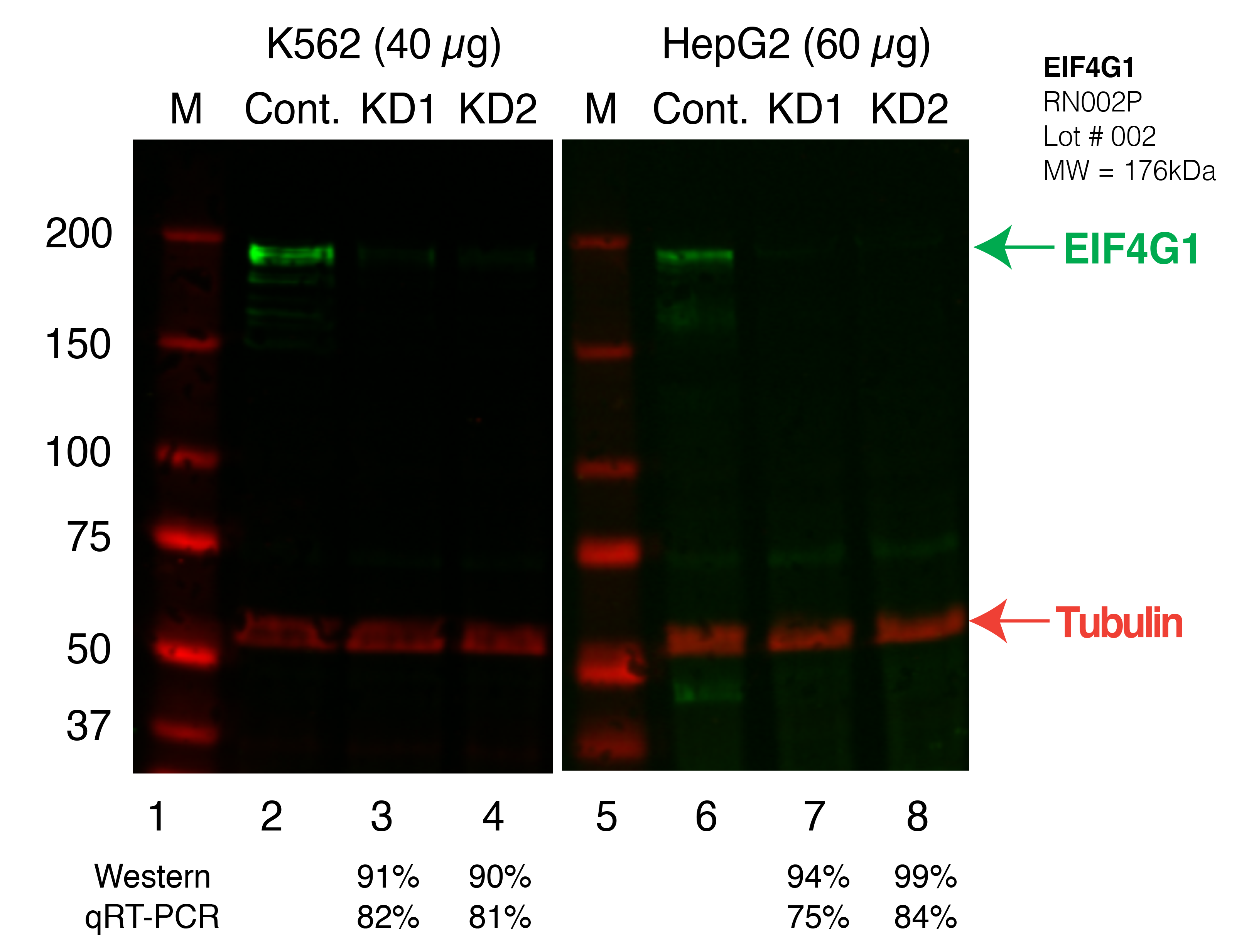
| 
|
| | |
|---|
| EIF4G1 | Product_ID: 8701S
Lot_ID: 1
Source: Cell Signaling Technology
Target Name: EIF4G1-human | | | 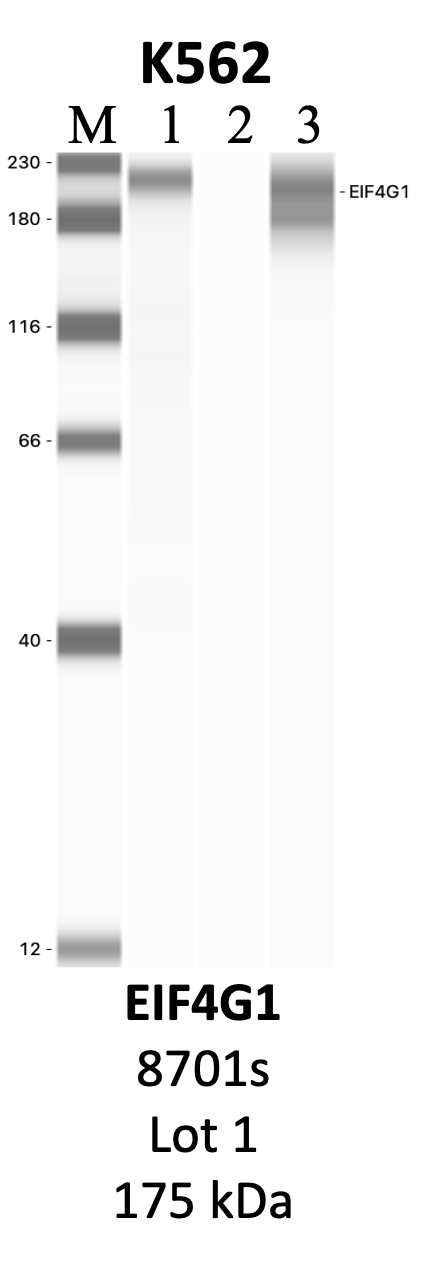
| | | |
|---|