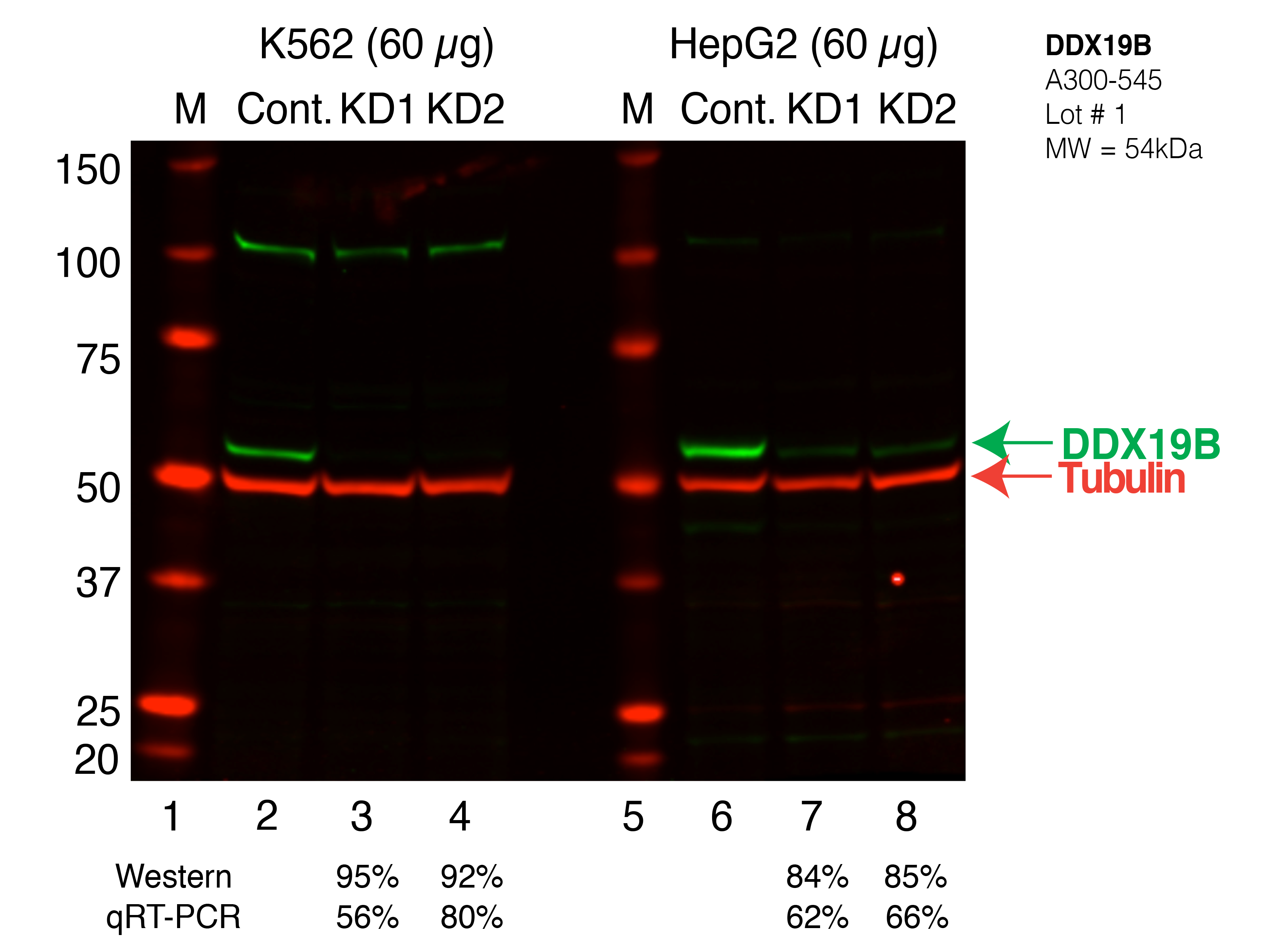
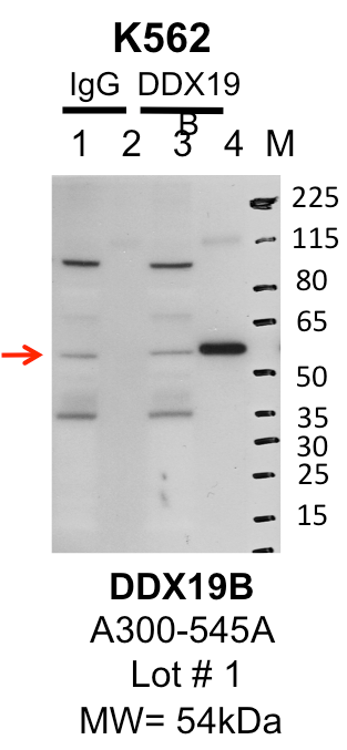
| DDX19B | Product_ID: A300-545A
Lot_ID: 1
Source: Bethyl Labs
Target Name: DDX19B-human | 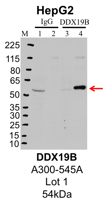
| 
| 
|
| | |
|---|
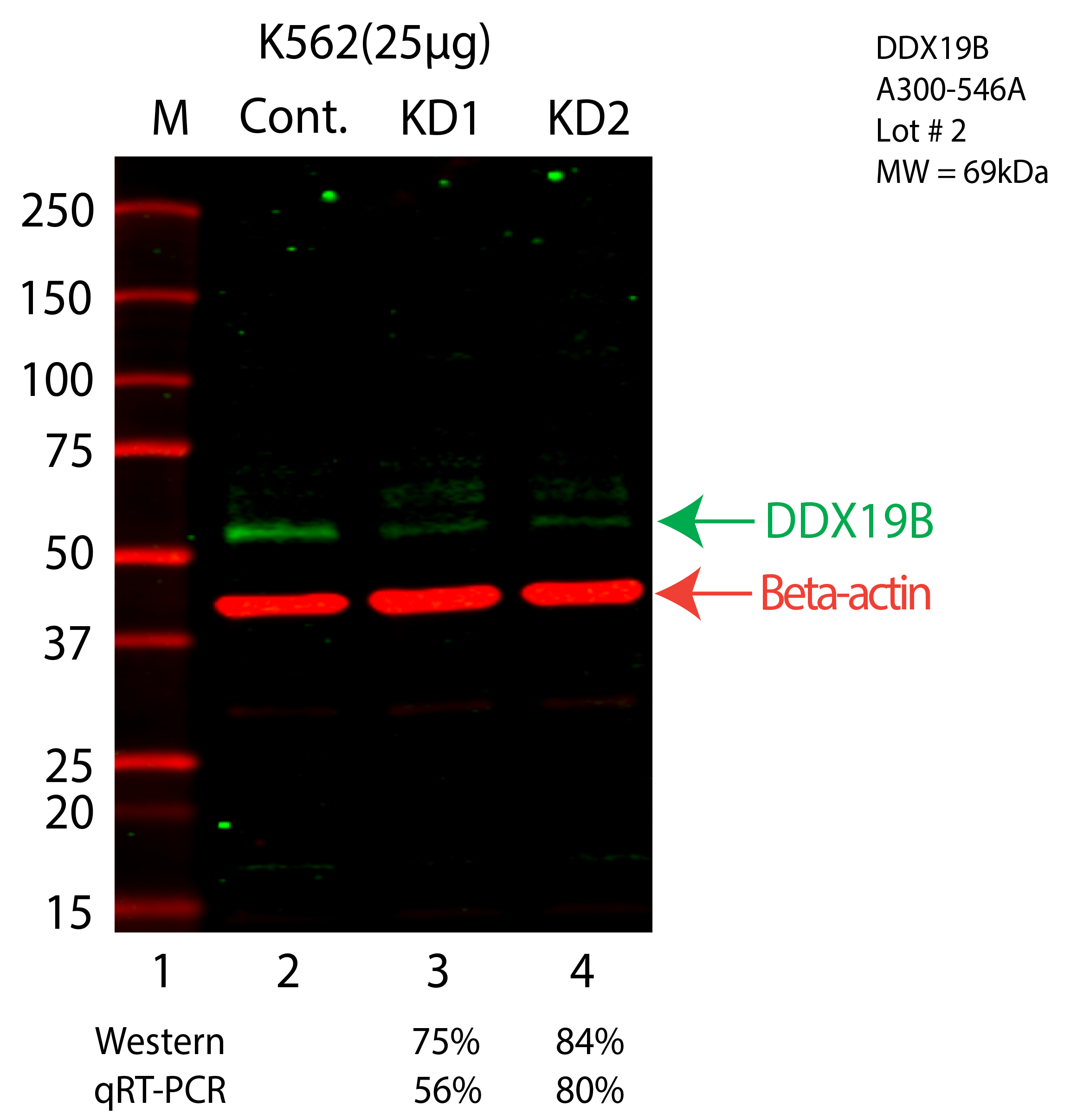
| DDX19B | Product_ID: A300-546A
Lot_ID: 1
Source: Bethyl Labs
Target Name: DDX19B-human | | | 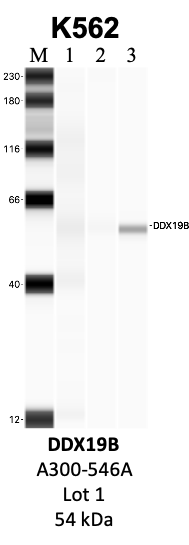
| 
| | |
|---|
| DDX19B | Product_ID: A300-547A
Lot_ID: 1
Source: Bethyl Labs
Target Name: DDX19B-human | | | 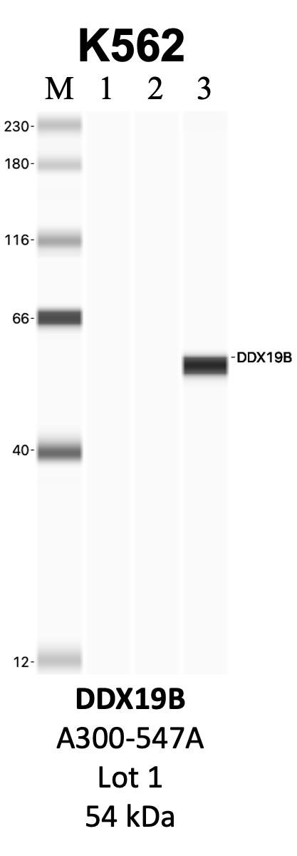
| | | |
|---|