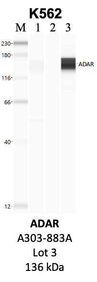
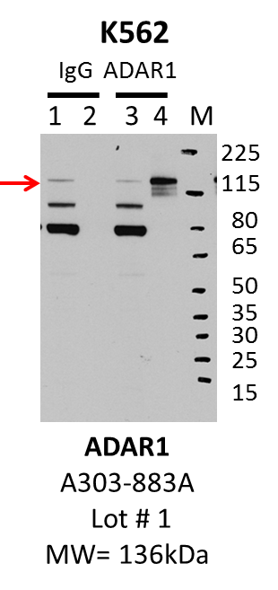
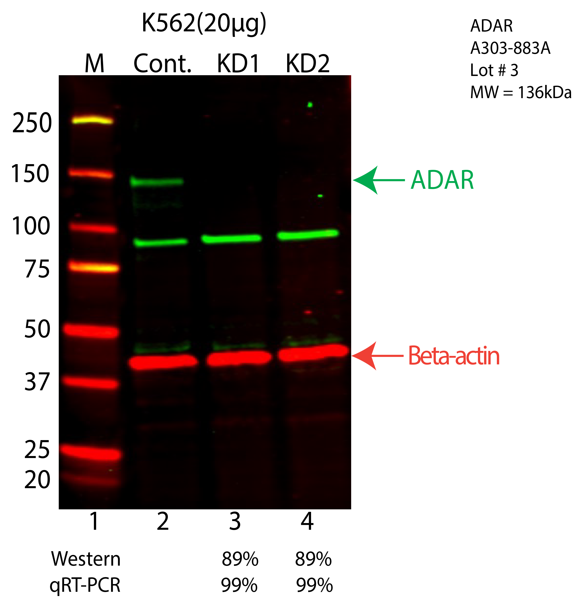
| ADAR | Product_ID: A303-883A
Lot_ID: 1
Source: Bethyl Labs
Target Name: ADAR-human | | 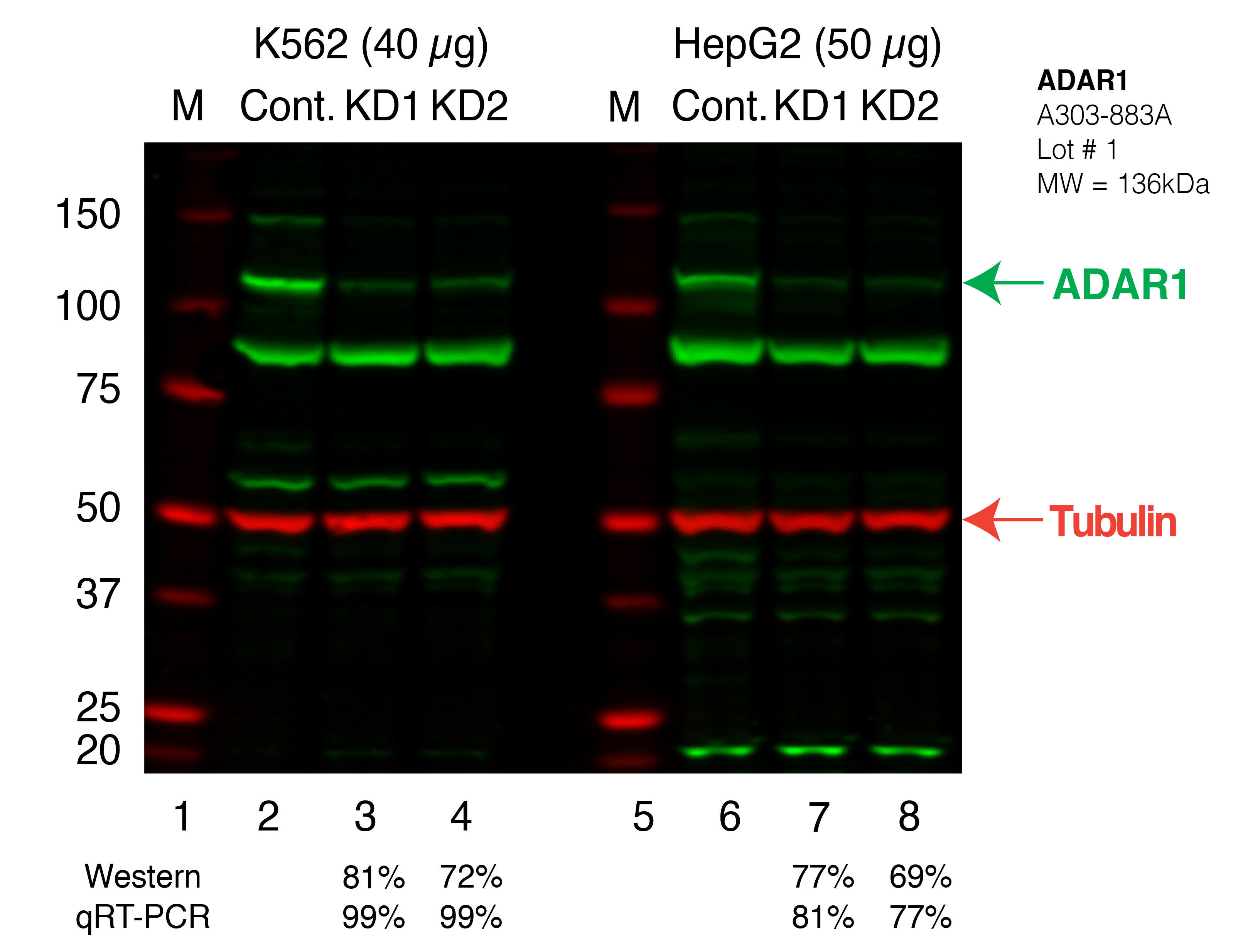
| 
|
| | |
|---|
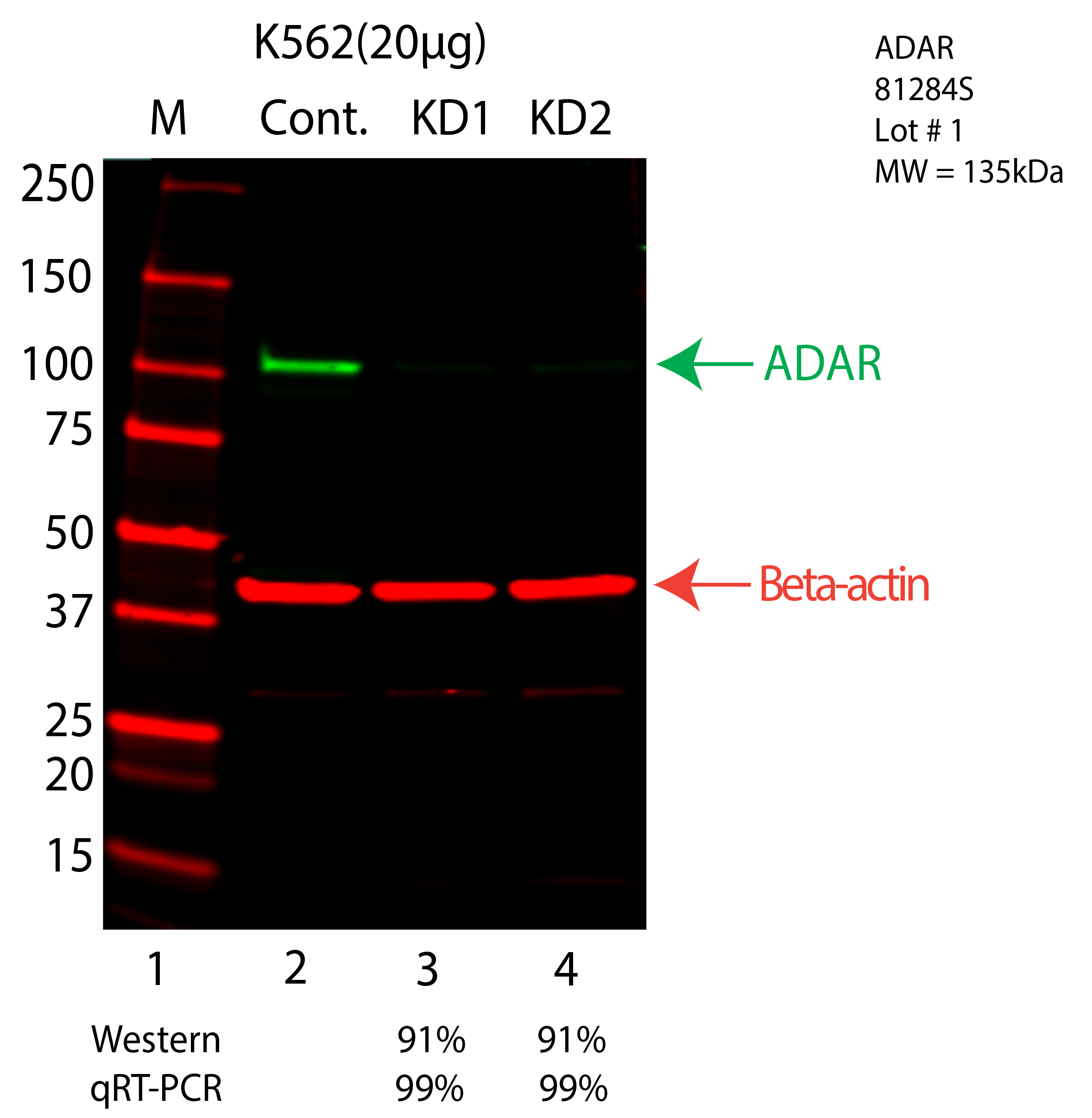
| ADAR | Product_ID: 81284S
Lot_ID: 1
Source: Cell Signaling Technology
Target Name: ADAR-human | | | 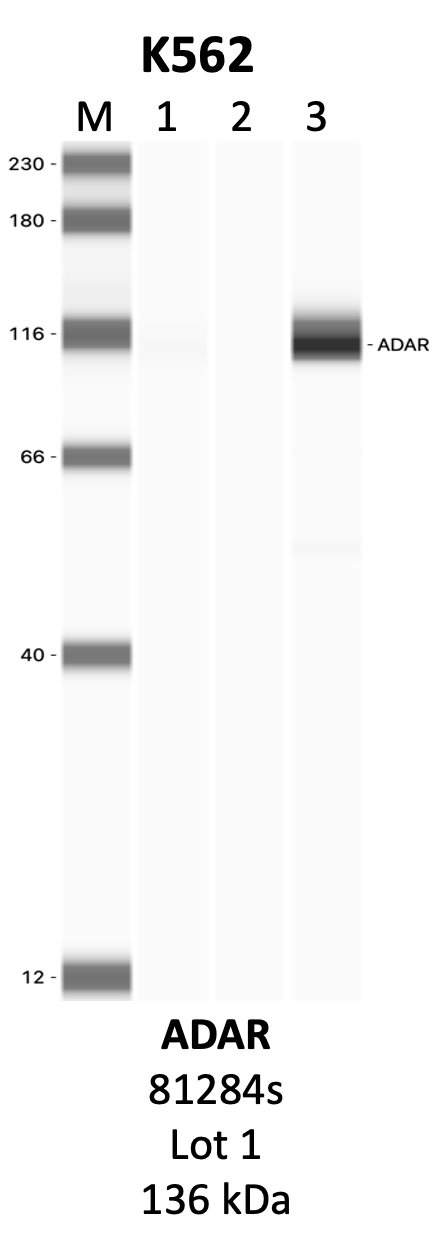
| 
| | |
|---|
| ADAR | Product_ID: SC-73408
Lot_ID: 1
Source: Santa Cruz Biotech
Target Name: ADAR-human | | | | 
| | |
|---|